 Movie
Movie Controller

[English] 日本語
 Yorodumi
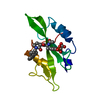
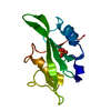
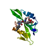
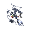
- PDB-1sha: CRYSTAL STRUCTURE OF THE PHOSPHOTYROSINE RECOGNITION DOMAIN SH2 O... -
+ Open data

- Basic information
| Entry | Database: PDB / ID: 1sha | ||||||
|---|---|---|---|---|---|---|---|
| Title | CRYSTAL STRUCTURE OF THE PHOSPHOTYROSINE RECOGNITION DOMAIN SH2 OF V-SRC COMPLEXED WITH TYROSINE-PHOSPHORYLATED PEPTIDES | ||||||
 Components Components |
| ||||||
 Keywords Keywords |  PHOSPHOTRANSFERASE PHOSPHOTRANSFERASE | ||||||
| Function / homology |  Function and homology information Function and homology informationplatelet activating factor receptor activity / platelet-derived growth factor receptor activity / platelet-derived growth factor beta-receptor activity / cell migration involved in coronary angiogenesis / metanephric glomerular mesangial cell proliferation involved in metanephros development / positive regulation of metanephric mesenchymal cell migration by platelet-derived growth factor receptor-beta signaling pathway / smooth muscle cell chemotaxis / metanephric glomerular capillary formation / cell migration involved in vasculogenesis / aorta morphogenesis ...platelet activating factor receptor activity / platelet-derived growth factor receptor activity / platelet-derived growth factor beta-receptor activity / cell migration involved in coronary angiogenesis / metanephric glomerular mesangial cell proliferation involved in metanephros development / positive regulation of metanephric mesenchymal cell migration by platelet-derived growth factor receptor-beta signaling pathway / smooth muscle cell chemotaxis / metanephric glomerular capillary formation / cell migration involved in vasculogenesis / aorta morphogenesis / positive regulation of cell proliferation by VEGF-activated platelet derived growth factor receptor signaling pathway / platelet-derived growth factor binding / retina vasculature development in camera-type eye / vascular endothelial growth factor binding / cardiac myofibril assembly / phosphatidylinositol metabolic process / positive regulation of chemotaxis / Signaling by PDGF / platelet-derived growth factor receptor binding / positive regulation of DNA biosynthetic process / positive regulation of smooth muscle cell migration / positive regulation of calcium ion import / platelet-derived growth factor receptor-beta signaling pathway / positive regulation of phosphoprotein phosphatase activity / platelet-derived growth factor receptor signaling pathway / positive regulation of phospholipase C activity / positive regulation of calcium-mediated signaling / cell chemotaxis / Downstream signal transduction / lysosomal lumen / positive regulation of mitotic nuclear division / regulation of actin cytoskeleton organization / positive regulation of smooth muscle cell proliferation / non-specific protein-tyrosine kinase / non-membrane spanning protein tyrosine kinase activity / positive regulation of MAP kinase activity / receptor protein-tyrosine kinase / peptidyl-tyrosine phosphorylation / cell surface receptor protein tyrosine kinase signaling pathway / Constitutive Signaling by Aberrant PI3K in Cancer / positive regulation of reactive oxygen species metabolic process / PIP3 activates AKT signaling / PI5P, PP2A and IER3 Regulate PI3K/AKT Signaling / cytoplasmic vesicle / RAF/MAP kinase cascade / protein tyrosine kinase activity / protein autophosphorylation / positive regulation of phosphatidylinositol 3-kinase/protein kinase B signal transduction / positive regulation of ERK1 and ERK2 cascade / receptor complex / protein kinase activity / positive regulation of cell migration / apical plasma membrane / phosphorylation / signaling receptor binding / focal adhesion / intracellular membrane-bounded organelle / positive regulation of cell population proliferation / protein kinase binding / Golgi apparatus / enzyme binding / signal transduction / ATP binding / membrane / nucleus / plasma membrane / cytoplasmSimilarity search - Function | ||||||
| Biological species |  Rous sarcoma virus Rous sarcoma virus | ||||||
| Method | X-RAY DIFFRACTION / Resolution: 1.5 Å | ||||||
 Authors Authors | Waksman, G. / Kuriyan, J. | ||||||
 Citation Citation | Journal: Nature / Year: 1992 Title: Crystal structure of the phosphotyrosine recognition domain SH2 of v-src complexed with tyrosine-phosphorylated peptides. Authors: Waksman, G. / Kominos, D. / Robertson, S.C. / Pant, N. / Baltimore, D. / Birge, R.B. / Cowburn, D. / Hanafusa, H. / Mayer, B.J. / Overduin, M. / Resh, M.D. / Rios, C.B. / Silverman, L. / Kuriyan, J. | ||||||
| History |
|
- Structure visualization
| Structure viewer | Molecule: MolmilJmol/JSmol |
|---|
- Downloads & links
-Download
| PDBx/mmCIF format | 1sha.cif.gz | 30.2 KB | Display | PDBx/mmCIF format |
|---|---|---|---|---|
| PDB format | pdb1sha.ent.gz | 23.7 KB | Display | PDB format |
| PDBx/mmJSON format | 1sha.json.gz | Tree view | PDBx/mmJSON format | |
| Others |  Other downloads Other downloads |
-Validation report
| Arichive directory | https://data.pdbj.org/pub/pdb/validation_reports/sh/1shaftp://data.pdbj.org/pub/pdb/validation_reports/sh/1sha | HTTPS FTP |
|---|
-Related structure data


-Links
 PDBj
PDBj


- Assembly
| Deposited unit | 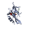
| ||||||||
|---|---|---|---|---|---|---|---|---|---|
| 1 |
| ||||||||
| Unit cell |
|
-Components
| #1: Protein | Mass: 11970.480 Da / Num. of mol.: 1 Source method: isolated from a genetically manipulated source Source: (gene. exp.) Rous sarcoma virus / Genus: Alpharetrovirus / References: UniProt: P00524, EC: 2.7.1.112 |
|---|---|
| #2: Protein/peptide | Mass: 701.767 Da / Num. of mol.: 1 Source method: isolated from a genetically manipulated source Source: (gene. exp.) Rous sarcoma virus / Genus: Alpharetrovirus / References: UniProt: P09619*PLUS |
| #3: Water | ChemComp-HOH / Water Mass: 18.015 Da / Num. of mol.: 58 / Source method: isolated from a natural source / Formula: H2O Mass: 18.015 Da / Num. of mol.: 58 / Source method: isolated from a natural source / Formula: H2O |
-Experimental details
-Experiment
| Experiment | Method: X-RAY DIFFRACTION |
|---|
- Sample preparation
| Crystal | Density Matthews: 2.08 Å3/Da / Density % sol: 40.77 % | ||||||||||||||||||||
|---|---|---|---|---|---|---|---|---|---|---|---|---|---|---|---|---|---|---|---|---|---|
| Crystal grow | *PLUS pH: 8 / Method: vapor diffusion | ||||||||||||||||||||
| Components of the solutions | *PLUS
|
- Processing
| Software |
| ||||||||||||||||||||||||||||||||||||||||||||||||||||||||||||
|---|---|---|---|---|---|---|---|---|---|---|---|---|---|---|---|---|---|---|---|---|---|---|---|---|---|---|---|---|---|---|---|---|---|---|---|---|---|---|---|---|---|---|---|---|---|---|---|---|---|---|---|---|---|---|---|---|---|---|---|---|---|
| Refinement | Resolution: 1.5→6 Å / Rfactor Rwork: 0.208 / Rfactor obs: 0.208 / σ(F): 2 Details: THE LOOP FROM RESIDUE 49 TO 53 IS DISORDERED AND IS NOT CLEARLY VISIBLE IN THE ELECTRON DENSITY. | ||||||||||||||||||||||||||||||||||||||||||||||||||||||||||||
| Refinement step | Cycle: LAST / Resolution: 1.5→6 Å
| ||||||||||||||||||||||||||||||||||||||||||||||||||||||||||||
| Refine LS restraints |
| ||||||||||||||||||||||||||||||||||||||||||||||||||||||||||||
| Software | *PLUS Name: X-PLOR / Classification: refinement | ||||||||||||||||||||||||||||||||||||||||||||||||||||||||||||
| Refinement | *PLUS Rfactor obs: 0.208 | ||||||||||||||||||||||||||||||||||||||||||||||||||||||||||||
| Solvent computation | *PLUS | ||||||||||||||||||||||||||||||||||||||||||||||||||||||||||||
| Displacement parameters | *PLUS | ||||||||||||||||||||||||||||||||||||||||||||||||||||||||||||
| Refine LS restraints | *PLUS Type: x_angle_d / Dev ideal: 2.9 |
