 Movie
Movie Controller

[English] 日本語
 Yorodumi
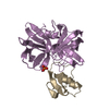
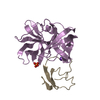
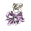
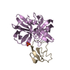
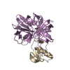
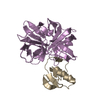
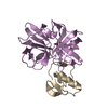
- PDB-1sgp: ALA 18 VARIANT OF TURKEY OVOMUCOID INHIBITOR THIRD DOMAIN COMPLEX... -
+ Open data

- Basic information
| Entry | Database: PDB / ID: 1sgp | ||||||
|---|---|---|---|---|---|---|---|
| Title | ALA 18 VARIANT OF TURKEY OVOMUCOID INHIBITOR THIRD DOMAIN COMPLEXED WITH STREPTOMYCES GRISEUS PROTEINASE B | ||||||
 Components Components |
| ||||||
 Keywords Keywords | COMPLEX (SERINE PROTEASE/INHIBITOR) /  SERINE PROTEINASE / PROTEIN INHIBITOR / COMPLEX (SERINE PROTEASE-INHIBITOR) COMPLEX SERINE PROTEINASE / PROTEIN INHIBITOR / COMPLEX (SERINE PROTEASE-INHIBITOR) COMPLEX | ||||||
| Function / homology |  Function and homology informationstreptogrisin B / molecular function inhibitor activity / serine-type endopeptidase inhibitor activity / serine-type endopeptidase activity / proteolysis / extracellular region Function and homology informationstreptogrisin B / molecular function inhibitor activity / serine-type endopeptidase inhibitor activity / serine-type endopeptidase activity / proteolysis / extracellular regionSimilarity search - Function | ||||||
| Biological species |  Streptomyces griseus (bacteria) Streptomyces griseus (bacteria) Meleagris gallopavo (turkey) Meleagris gallopavo (turkey) | ||||||
| Method | X-RAY DIFFRACTION / SYNCHROTRON / Resolution: 1.4 Å | ||||||
 Authors Authors | Huang, K. / James, M.N.G. | ||||||
 Citation Citation | Journal: Protein Sci. / Year: 1995 Title: Water molecules participate in proteinase-inhibitor interactions: crystal structures of Leu18, Ala18, and Gly18 variants of turkey ovomucoid inhibitor third domain complexed with Streptomyces griseus proteinase B. Authors: Huang, K. / Lu, W. / Anderson, S. / Laskowski Jr., M. / James, M.N. #1: Journal: Biochemistry / Year: 1983Title: Structure of the Complex of Streptomyces Griseus Protease B and the Third Domain of the Turkey Ovomucoid Inhibitor at 1.8 Angstroms Resolution Authors: Read, R.J. / Fujinaga, M. / Sielecki, A.R. / James, M.N.G. | ||||||
| History |
|
- Structure visualization
| Structure viewer | Molecule: MolmilJmol/JSmol |
|---|
- Downloads & links
-Download
| PDBx/mmCIF format | 1sgp.cif.gz | 56 KB | Display | PDBx/mmCIF format |
|---|---|---|---|---|
| PDB format | pdb1sgp.ent.gz | 42.6 KB | Display | PDB format |
| PDBx/mmJSON format | 1sgp.json.gz | Tree view | PDBx/mmJSON format | |
| Others |  Other downloads Other downloads |
-Validation report
| Arichive directory | https://data.pdbj.org/pub/pdb/validation_reports/sg/1sgpftp://data.pdbj.org/pub/pdb/validation_reports/sg/1sgp | HTTPS FTP |
|---|
-Related structure data


-Links
 PDBj
PDBj
- Assembly
| Deposited unit | 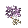
| ||||||||
|---|---|---|---|---|---|---|---|---|---|
| 1 |
| ||||||||
| Unit cell |
| ||||||||
| Atom site foot note | 1: CIS PROLINE - PRO E 99A / 2: CIS PROLINE - PRO I 12 |
-Components
| #1: Protein | / SGPB Mass: 18665.285 Da / Num. of mol.: 1 Source method: isolated from a genetically manipulated source Source: (gene. exp.) Streptomyces griseus (bacteria) / Strain: K1 / Plasmid: PEZZ318.TKY / Production host: Escherichia coli (E. coli) / References: UniProt: P00777, streptogrisin B |
|---|---|
| #2: Protein | Mass: 5543.208 Da / Num. of mol.: 1 / Mutation: DEL(1-6), L18A Source method: isolated from a genetically manipulated source Source: (gene. exp.) Meleagris gallopavo (turkey) / Plasmid: PEZZ318.TKY / Production host: Escherichia coli (E. coli) / References: UniProt: P68390 |
| #3: Chemical | ChemComp-PO4 / Phosphate  Mass: 94.971 Da / Num. of mol.: 1 / Source method: obtained synthetically / Formula: PO4 Mass: 94.971 Da / Num. of mol.: 1 / Source method: obtained synthetically / Formula: PO4 |
| #4: Water | ChemComp-HOH / Water Mass: 18.015 Da / Num. of mol.: 181 / Source method: isolated from a natural source / Formula: H2O Mass: 18.015 Da / Num. of mol.: 181 / Source method: isolated from a natural source / Formula: H2O |
-Experimental details
-Experiment
| Experiment | Method: X-RAY DIFFRACTION |
|---|
- Sample preparation
| Crystal | Density Matthews: 2.03 Å3/Da / Density % sol: 39.45 % | ||||||||||||||||||||||||||||||
|---|---|---|---|---|---|---|---|---|---|---|---|---|---|---|---|---|---|---|---|---|---|---|---|---|---|---|---|---|---|---|---|
| Crystal grow | *PLUS pH: 6.5 / Method: vapor diffusion, hanging drop / Details: used to seeding | ||||||||||||||||||||||||||||||
| Components of the solutions | *PLUS
|
-Data collection
| Diffraction source | Source: SYNCHROTRON / Site: Photon Factory  / Beamline: BL-18B / Wavelength: 1 / Beamline: BL-18B / Wavelength: 1 |
|---|---|
| Detector | Type: WEISSENBERG / Detector: DIFFRACTOMETER / Date: Nov 28, 1993 |
| Radiation | Monochromatic (M) / Laue (L): M / Scattering type: x-ray |
| Radiation wavelength | Wavelength: 1 Å / Relative weight: 1 |
| Reflection | Redundancy: 2.7 % / Rmerge(I) obs: 0.0733 |
| Reflection | *PLUS Highest resolution: 1.4 Å / Num. obs: 30783 / % possible obs: 80.3 % / Num. measured all: 83250 / Rmerge(I) obs: 0.0733 |
| Reflection shell | *PLUS Highest resolution: 1.4 Å / Lowest resolution: 1.5 Å / % possible obs: 56 % |
- Processing
| Software |
| ||||||||||||||||||||||||||||||
|---|---|---|---|---|---|---|---|---|---|---|---|---|---|---|---|---|---|---|---|---|---|---|---|---|---|---|---|---|---|---|---|
| Refinement | Resolution: 1.4→20 Å / σ(F): 1 /
| ||||||||||||||||||||||||||||||
| Refinement step | Cycle: LAST / Resolution: 1.4→20 Å
| ||||||||||||||||||||||||||||||
| Refine LS restraints |
| ||||||||||||||||||||||||||||||
| Software | *PLUS Name: TNT / Classification: refinement | ||||||||||||||||||||||||||||||
| Refine LS restraints | *PLUS
|
