 Movie
Movie Controller


+ Open data

- Basic information
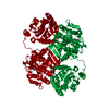
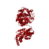
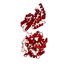
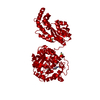
| Entry | Database: PDB / ID: 1s8o | |||||||||
|---|---|---|---|---|---|---|---|---|---|---|
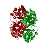
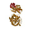
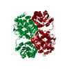
| Title | Human soluble Epoxide Hydrolase | |||||||||
 Components Components | epoxide hydrolase 2, cytoplasmic | |||||||||
 Keywords Keywords | HYDROLASE / domain-swapped dimer | |||||||||
| Function / homology |  Function and homology informationlipid-phosphate phosphatase / 10-hydroxy-9-(phosphonooxy)octadecanoate phosphatase activity / stilbene catabolic process / Biosynthesis of maresins / phospholipid dephosphorylation / soluble epoxide hydrolase / lipid phosphatase activity / epoxide metabolic process / lysophosphatidic acid phosphatase activity / Synthesis of epoxy (EET) and dihydroxyeicosatrienoic acids (DHET) ...lipid-phosphate phosphatase / 10-hydroxy-9-(phosphonooxy)octadecanoate phosphatase activity / stilbene catabolic process / Biosynthesis of maresins / phospholipid dephosphorylation / soluble epoxide hydrolase / lipid phosphatase activity / epoxide metabolic process / lysophosphatidic acid phosphatase activity / Synthesis of epoxy (EET) and dihydroxyeicosatrienoic acids (DHET) / epoxide hydrolase activity / regulation of cholesterol metabolic process / phosphatase activity / peroxisomal matrix / toxic substance binding / dephosphorylation / cholesterol homeostasis / Peroxisomal protein import / response to toxic substance / peroxisome / positive regulation of gene expression / magnesium ion binding / protein homodimerization activity / extracellular exosome / cytosol Function and homology informationlipid-phosphate phosphatase / 10-hydroxy-9-(phosphonooxy)octadecanoate phosphatase activity / stilbene catabolic process / Biosynthesis of maresins / phospholipid dephosphorylation / soluble epoxide hydrolase / lipid phosphatase activity / epoxide metabolic process / lysophosphatidic acid phosphatase activity / Synthesis of epoxy (EET) and dihydroxyeicosatrienoic acids (DHET) ...lipid-phosphate phosphatase / 10-hydroxy-9-(phosphonooxy)octadecanoate phosphatase activity / stilbene catabolic process / Biosynthesis of maresins / phospholipid dephosphorylation / soluble epoxide hydrolase / lipid phosphatase activity / epoxide metabolic process / lysophosphatidic acid phosphatase activity / Synthesis of epoxy (EET) and dihydroxyeicosatrienoic acids (DHET) / epoxide hydrolase activity / regulation of cholesterol metabolic process / phosphatase activity / peroxisomal matrix / toxic substance binding / dephosphorylation / cholesterol homeostasis / Peroxisomal protein import / response to toxic substance / peroxisome / positive regulation of gene expression / magnesium ion binding / protein homodimerization activity / extracellular exosome / cytosolSimilarity search - Function | |||||||||
| Biological species |  Homo sapiens (human) Homo sapiens (human) | |||||||||
| Method | X-RAY DIFFRACTION / SYNCHROTRON / MOLECULAR REPLACEMENT / Resolution: 2.6 Å | |||||||||
 Authors Authors | Gomez, G.A. / Morisseau, C. / Hammock, B.D. / Christianson, D.W. | |||||||||
 Citation Citation | Journal: Biochemistry / Year: 2004 Title: Structure of human epoxide hydrolase reveals mechanistic inferences on bifunctional catalysis in epoxide and phosphate ester hydrolysis Authors: Gomez, G.A. / Morisseau, C. / Hammock, B.D. / Christianson, D.W. | |||||||||
| History |
| |||||||||
| Remark 999 | SEQUENCE The numbering of amino acids was modified to adopt the numbering employed in murine ...SEQUENCE The numbering of amino acids was modified to adopt the numbering employed in murine epoxide hydrolase (pdb entry 1CQZ). |
- Structure visualization
| Structure viewer | Molecule: MolmilJmol/JSmol |
|---|
- Downloads & links
-Download
| PDBx/mmCIF format | 1s8o.cif.gz | 120.4 KB | Display | PDBx/mmCIF format |
|---|---|---|---|---|
| PDB format | pdb1s8o.ent.gz | 93.4 KB | Display | PDB format |
| PDBx/mmJSON format | 1s8o.json.gz | Tree view | PDBx/mmJSON format | |
| Others |  Other downloads Other downloads |
-Validation report
| Arichive directory | https://data.pdbj.org/pub/pdb/validation_reports/s8/1s8oftp://data.pdbj.org/pub/pdb/validation_reports/s8/1s8o | HTTPS FTP |
|---|
-Related structure data
| Related structure data | 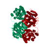 1vj5C 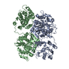 1cqzS C: citing same article ( S: Starting model for refinement |
|---|---|
| Similar structure data |


-Links
 PDBj
PDBj


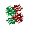
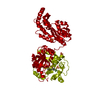
- Assembly
| Deposited unit | 
| ||||||||
|---|---|---|---|---|---|---|---|---|---|
| 1 | 
| ||||||||
| Unit cell |
| ||||||||
| Components on special symmetry positions |
| ||||||||
| Details | The second part of the biological dimer is assembeled by the crystollographic two fold axis. |
-Components
| #1: Protein | / HsEH Mass: 62685.617 Da / Num. of mol.: 1 Source method: isolated from a genetically manipulated source Source: (gene. exp.) Homo sapiens (human) / Gene: EPHX2 / Cell line (production host): High Five / Production host:   Spodoptera frugiperda (fall armyworm) / References: UniProt: P34913, epoxide hydrolase Spodoptera frugiperda (fall armyworm) / References: UniProt: P34913, epoxide hydrolase |
|---|---|
| #2: Chemical | ChemComp-P6G / Polyethylene glycol  Mass: 282.331 Da / Num. of mol.: 1 / Source method: obtained synthetically / Formula: C12H26O7 / Comment: precipitant*YM Mass: 282.331 Da / Num. of mol.: 1 / Source method: obtained synthetically / Formula: C12H26O7 / Comment: precipitant*YM |
| #3: Water | ChemComp-HOH / Water Mass: 18.015 Da / Num. of mol.: 46 / Source method: isolated from a natural source / Formula: H2O Mass: 18.015 Da / Num. of mol.: 46 / Source method: isolated from a natural source / Formula: H2O |
-Experimental details
-Experiment
| Experiment | Method: X-RAY DIFFRACTION / Number of used crystals: 1 |
|---|
- Sample preparation
| Crystal | Density Matthews: 2.41 Å3/Da / Density % sol: 48.98 % |
|---|---|
| Crystal grow | Temperature: 277 K / Method: vapor diffusion / pH: 8.4 Details: PEG 4000, Tris, lithium sulfate, pH 8.4, VAPOR DIFFUSION, temperature 277K |
-Data collection
| Diffraction | Mean temperature: 100 K |
|---|---|
| Diffraction source | Source: SYNCHROTRON / Site: CHESS  / Beamline: F1 / Wavelength: 0.9 Å / Beamline: F1 / Wavelength: 0.9 Å |
| Detector | Type: ADSC QUANTUM 4 / Detector: CCD / Date: Jan 18, 2002 |
| Radiation | Monochromator: Si111 / Protocol: SINGLE WAVELENGTH / Monochromatic (M) / Laue (L): M / Scattering type: x-ray |
| Radiation wavelength | Wavelength: 0.9 Å / Relative weight: 1 |
| Reflection | Resolution: 2.6→30 Å / Num. all: 19912 / Num. obs: 19879 / % possible obs: 99.83 % / Observed criterion σ(I): -3 / Redundancy: 9.9 % / Biso Wilson estimate: 37.1 Å2 / Rmerge(I) obs: 0.1 / Net I/σ(I): 30.9 |
| Reflection shell | Resolution: 2.6→2.69 Å / Rmerge(I) obs: 0.363 / Mean I/σ(I) obs: 11.5 / % possible all: 99.9 |
- Processing
| Software |
| ||||||||||||||||||||||||||||||||||||
|---|---|---|---|---|---|---|---|---|---|---|---|---|---|---|---|---|---|---|---|---|---|---|---|---|---|---|---|---|---|---|---|---|---|---|---|---|---|
| Refinement | Method to determine structure: MOLECULAR REPLACEMENT Starting model: PDB ENTRY 1CQZ Resolution: 2.6→29.41 Å / Rfactor Rfree error: 0.007 / Data cutoff high absF: 251057.73 / Data cutoff low absF: 0 / Isotropic thermal model: RESTRAINED / Cross valid method: THROUGHOUT / σ(F): 0 / Stereochemistry target values: Engh & Huber
| ||||||||||||||||||||||||||||||||||||
| Solvent computation | Solvent model: FLAT MODEL / Bsol: 26.355 Å2 / ksol: 0.359269 e/Å3 | ||||||||||||||||||||||||||||||||||||
| Displacement parameters | Biso mean: 34.7 Å2
| ||||||||||||||||||||||||||||||||||||
| Refine analyze |
| ||||||||||||||||||||||||||||||||||||
| Refinement step | Cycle: LAST / Resolution: 2.6→29.41 Å
| ||||||||||||||||||||||||||||||||||||
| Refine LS restraints |
| ||||||||||||||||||||||||||||||||||||
| LS refinement shell | Resolution: 2.6→2.76 Å / Rfactor Rfree error: 0.021 / Total num. of bins used: 6
| ||||||||||||||||||||||||||||||||||||
| Xplor file |
|
