 Movie
Movie Controller

[English] 日本語
 Yorodumi
- PDB-1s5p: Structure and substrate binding properties of cobB, a Sir2 homolo... -
+ Open data

- Basic information
| Entry | Database: PDB / ID: 1s5p | ||||||
|---|---|---|---|---|---|---|---|
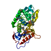
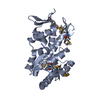
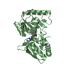
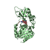
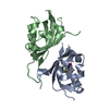
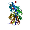
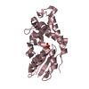
| Title | Structure and substrate binding properties of cobB, a Sir2 homolog protein deacetylase from Eschericia coli. | ||||||
 Components Components |
| ||||||
 Keywords Keywords |  HYDROLASE / protein deacetylase / Sir2 homologue HYDROLASE / protein deacetylase / Sir2 homologue | ||||||
| Function / homology |  Function and homology information Function and homology informationlipoamidase activity / NAD-dependent protein de-2-hydroxyisobutyrylase activity / protein-malonyllysine demalonylase activity / protein-succinyllysine desuccinylase activity / NAD-dependent protein lysine deacetylase activity / cyclic-di-GMP binding / protein acetyllysine N-acetyltransferase / NAD-dependent histone deacetylase activity / NAD+ ADP-ribosyltransferase activity / NAD+ binding ...lipoamidase activity / NAD-dependent protein de-2-hydroxyisobutyrylase activity / protein-malonyllysine demalonylase activity / protein-succinyllysine desuccinylase activity / NAD-dependent protein lysine deacetylase activity / cyclic-di-GMP binding / protein acetyllysine N-acetyltransferase / NAD-dependent histone deacetylase activity / NAD+ ADP-ribosyltransferase activity / NAD+ binding / chemotaxis / defense response to virus / protein homodimerization activity / zinc ion binding / cytoplasmSimilarity search - Function | ||||||
| Biological species |  Escherichia coli (E. coli) Escherichia coli (E. coli) | ||||||
| Method | X-RAY DIFFRACTION / SYNCHROTRON / MAD / Resolution: 1.96 Å | ||||||
 Authors Authors | Zhao, K. / Chai, X. / Marmorstein, R. | ||||||
 Citation Citation | Journal: J.Mol.Biol. / Year: 2004 Title: Structure and Substrate Binding Properties of cobB, a Sir2 Homolog Protein Deacetylase from Eschericia coli. Authors: Zhao, K. / Chai, X. / Marmorstein, R. | ||||||
| History |
|
- Structure visualization
| Structure viewer | Molecule: MolmilJmol/JSmol |
|---|
- Downloads & links
-Download
| PDBx/mmCIF format | 1s5p.cif.gz | 60.7 KB | Display | PDBx/mmCIF format |
|---|---|---|---|---|
| PDB format | pdb1s5p.ent.gz | 47.5 KB | Display | PDB format |
| PDBx/mmJSON format | 1s5p.json.gz | Tree view | PDBx/mmJSON format | |
| Others |  Other downloads Other downloads |
-Validation report
| Arichive directory | https://data.pdbj.org/pub/pdb/validation_reports/s5/1s5pftp://data.pdbj.org/pub/pdb/validation_reports/s5/1s5p | HTTPS FTP |
|---|
-Related structure data
| Similar structure data |
|---|


-Links
 PDBj
PDBj

- Assembly
| Deposited unit | 
| ||||||||
|---|---|---|---|---|---|---|---|---|---|
| 1 |
| ||||||||
| Unit cell |
|
-Components
| #1: Protein | Mass: 26088.607 Da / Num. of mol.: 1 Source method: isolated from a genetically manipulated source Source: (gene. exp.) Escherichia coli (E. coli) / Gene: NPDA, COBB, B1120 / Species (production host): Escherichia coli / Production host: Escherichia coli BL21(DE3) (bacteria) / Strain (production host): bl21(de3)References: UniProt: P75960, Hydrolases; Acting on carbon-nitrogen bonds, other than peptide bonds; In linear amides |
|---|---|
| #2: Protein/peptide | Mass: 955.120 Da / Num. of mol.: 1 / Source method: obtained synthetically / Details: synthetic |
| #3: Chemical | ChemComp-ZN /   Mass: 65.409 Da / Num. of mol.: 1 / Source method: obtained synthetically / Formula: Zn Mass: 65.409 Da / Num. of mol.: 1 / Source method: obtained synthetically / Formula: Zn |
| #4: Water | ChemComp-HOH / Water Mass: 18.015 Da / Num. of mol.: 229 / Source method: isolated from a natural source / Formula: H2O Mass: 18.015 Da / Num. of mol.: 229 / Source method: isolated from a natural source / Formula: H2O |
-Experimental details
-Experiment
| Experiment | Method: X-RAY DIFFRACTION / Number of used crystals: 1 |
|---|
- Sample preparation
| Crystal | Density Matthews: 2.67 Å3/Da / Density % sol: 53.56 % | ||||||||||||||||||||||||||||||
|---|---|---|---|---|---|---|---|---|---|---|---|---|---|---|---|---|---|---|---|---|---|---|---|---|---|---|---|---|---|---|---|
| Crystal grow | *PLUS pH: 6.5 / Method: vapor diffusion, hanging drop | ||||||||||||||||||||||||||||||
| Components of the solutions | *PLUS
|
-Data collection
| Diffraction |
| |||||||||||||||
|---|---|---|---|---|---|---|---|---|---|---|---|---|---|---|---|---|
| Diffraction source |
| |||||||||||||||
| Radiation |
| |||||||||||||||
| Radiation wavelength |
| |||||||||||||||
| Reflection | Resolution: 1.96→50 Å / Num. obs: 20061 / % possible obs: 98.7 % / Observed criterion σ(F): 0 / Observed criterion σ(I): 0 / Redundancy: 8.1 % / Biso Wilson estimate: 29.6 Å2 / Rmerge(I) obs: 0.033 / Rsym value: 0.034 / Net I/σ(I): 55.5 | |||||||||||||||
| Reflection shell | Resolution: 1.96→2.03 Å / Rmerge(I) obs: 0.269 / Mean I/σ(I) obs: 6.4 / Num. unique all: 1950 / Rsym value: 0.244 / % possible all: 98.6 | |||||||||||||||
| Reflection shell | *PLUS % possible obs: 98 % / Rmerge(I) obs: 0.066 |

- Processing
| Software |
| ||||||||||||||||||||||||||||||||||||||||||||||||||||||||||||||||||||||||||||||||
|---|---|---|---|---|---|---|---|---|---|---|---|---|---|---|---|---|---|---|---|---|---|---|---|---|---|---|---|---|---|---|---|---|---|---|---|---|---|---|---|---|---|---|---|---|---|---|---|---|---|---|---|---|---|---|---|---|---|---|---|---|---|---|---|---|---|---|---|---|---|---|---|---|---|---|---|---|---|---|---|---|---|
| Refinement | Method to determine structure: MAD / Resolution: 1.96→29.7 Å / Rfactor Rfree error: 0.007 / Data cutoff high absF: 928333.29 / Data cutoff low absF: 0 / Isotropic thermal model: RESTRAINED / Cross valid method: THROUGHOUT / σ(F): 0 / σ(I): 0 / Stereochemistry target values: Engh & Huber
| ||||||||||||||||||||||||||||||||||||||||||||||||||||||||||||||||||||||||||||||||
| Solvent computation | Solvent model: FLAT MODEL / Bsol: 77.6041 Å2 / ksol: 0.375912 e/Å3 | ||||||||||||||||||||||||||||||||||||||||||||||||||||||||||||||||||||||||||||||||
| Displacement parameters | Biso mean: 49.9 Å2
| ||||||||||||||||||||||||||||||||||||||||||||||||||||||||||||||||||||||||||||||||
| Refine analyze |
| ||||||||||||||||||||||||||||||||||||||||||||||||||||||||||||||||||||||||||||||||
| Refinement step | Cycle: LAST / Resolution: 1.96→29.7 Å
| ||||||||||||||||||||||||||||||||||||||||||||||||||||||||||||||||||||||||||||||||
| Refine LS restraints |
| ||||||||||||||||||||||||||||||||||||||||||||||||||||||||||||||||||||||||||||||||
| LS refinement shell | Resolution: 1.96→2.08 Å / Rfactor Rfree error: 0.026 / Total num. of bins used: 6
| ||||||||||||||||||||||||||||||||||||||||||||||||||||||||||||||||||||||||||||||||
| Xplor file |
| ||||||||||||||||||||||||||||||||||||||||||||||||||||||||||||||||||||||||||||||||
| Refinement | *PLUS Lowest resolution: 30 Å / % reflection Rfree: 7 % / Rfactor Rfree: 0.27 | ||||||||||||||||||||||||||||||||||||||||||||||||||||||||||||||||||||||||||||||||
| Solvent computation | *PLUS | ||||||||||||||||||||||||||||||||||||||||||||||||||||||||||||||||||||||||||||||||
| Displacement parameters | *PLUS | ||||||||||||||||||||||||||||||||||||||||||||||||||||||||||||||||||||||||||||||||
| Refine LS restraints | *PLUS
|
