 Movie
Movie Controller


+ Open data

- Basic information
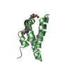
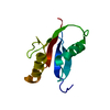
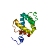
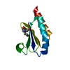
| Entry | Database: PDB / ID: 1s29 | ||||||
|---|---|---|---|---|---|---|---|
| Title | La autoantigen N-terminal domain | ||||||
 Components Components | La protein Sjögren syndrome antigen B Sjögren syndrome antigen B | ||||||
 Keywords Keywords | RNA BINDING PROTEIN / winged helix-turn-helix / autoantigen / RNA-binding | ||||||
| Function / homology |  Function and homology information Function and homology informationmaturation of 5S rRNA / tRNA processing / RNA processing / ribosome biogenesis / ribonucleoprotein complex / mRNA binding / RNA binding / nucleoplasm / nucleus / cytoplasmSimilarity search - Function | ||||||
| Biological species |  Trypanosoma brucei (eukaryote) Trypanosoma brucei (eukaryote) | ||||||
| Method | X-RAY DIFFRACTION / SYNCHROTRON / SAD / Resolution: 1.6 Å | ||||||
 Authors Authors | Dong, G. / Chakshusmathi, G. / Wolin, S.L. / Reinisch, K.M. | ||||||
 Citation Citation | Journal: Embo J. / Year: 2004 Title: Structure of the La motif: a winged helix domain mediates RNA binding via a conserved aromatic patch. Authors: Dong, G. / Chakshusmathi, G. / Wolin, S.L. / Reinisch, K.M. | ||||||
| History |
|
- Structure visualization
| Structure viewer | Molecule: MolmilJmol/JSmol |
|---|
- Downloads & links
-Download
| PDBx/mmCIF format | 1s29.cif.gz | 28 KB | Display | PDBx/mmCIF format |
|---|---|---|---|---|
| PDB format | pdb1s29.ent.gz | 21.1 KB | Display | PDB format |
| PDBx/mmJSON format | 1s29.json.gz | Tree view | PDBx/mmJSON format | |
| Others |  Other downloads Other downloads |
-Validation report
| Arichive directory | https://data.pdbj.org/pub/pdb/validation_reports/s2/1s29ftp://data.pdbj.org/pub/pdb/validation_reports/s2/1s29 | HTTPS FTP |
|---|
-Related structure data
| Related structure data | |
|---|---|
| Similar structure data |


-Links
 PDBj
PDBj
- Assembly
| Deposited unit | 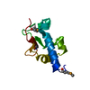
| ||||||||
|---|---|---|---|---|---|---|---|---|---|
| 1 |
| ||||||||
| Unit cell |
|
-Components
| #1: Protein | Sjögren syndrome antigen B Mass: 10651.736 Da / Num. of mol.: 1 / Fragment: N-terminal fragment Source method: isolated from a genetically manipulated source Source: (gene. exp.) Trypanosoma brucei (eukaryote) / Production host:  Escherichia coli (E. coli) / References: UniProt: Q9NIH4 Escherichia coli (E. coli) / References: UniProt: Q9NIH4 |
|---|---|
| #2: Water | ChemComp-HOH / Water Mass: 18.015 Da / Num. of mol.: 100 / Source method: isolated from a natural source / Formula: H2O Mass: 18.015 Da / Num. of mol.: 100 / Source method: isolated from a natural source / Formula: H2O |
-Experimental details
-Experiment
| Experiment | Method: X-RAY DIFFRACTION / Number of used crystals: 1 |
|---|
- Sample preparation
| Crystal | Density Matthews: 2.24 Å3/Da / Density % sol: 45.14 % / Description: Friedel pairs were used. | ||||||||||||||||||||||||||||||||||||
|---|---|---|---|---|---|---|---|---|---|---|---|---|---|---|---|---|---|---|---|---|---|---|---|---|---|---|---|---|---|---|---|---|---|---|---|---|---|
| Crystal grow | Temperature: 277 K / Method: vapor diffusion, hanging drop / pH: 7.5 Details: PEG3500, GLYCEROL, HEPES, DTT, pH 7.5, VAPOR DIFFUSION, HANGING DROP, temperature 277K | ||||||||||||||||||||||||||||||||||||
| Crystal grow | *PLUS Temperature: 4 ℃ / Method: vapor diffusion, hanging drop | ||||||||||||||||||||||||||||||||||||
| Components of the solutions | *PLUS
|
-Data collection
| Diffraction | Mean temperature: 110 K |
|---|---|
| Diffraction source | Source: SYNCHROTRON / Site: NSLS  / Beamline: X12C / Wavelength: 0.97 Å / Beamline: X12C / Wavelength: 0.97 Å |
| Detector | Type: BRANDEIS / Detector: CCD / Date: Aug 15, 2003 |
| Radiation | Monochromator: double-crystal monochromator Si(111), beam focused by a toroidal mirror Protocol: SAD / Monochromatic (M) / Laue (L): M / Scattering type: x-ray |
| Radiation wavelength | Wavelength: 0.97 Å / Relative weight: 1 |
| Reflection | Resolution: 1.6→20 Å / Num. all: 24632 / Num. obs: 24632 / % possible obs: 99.5 % / Observed criterion σ(F): 0 / Observed criterion σ(I): 0 / Biso Wilson estimate: 15.4 Å2 / Rmerge(I) obs: 0.032 / Net I/σ(I): 41 |
| Reflection shell | Resolution: 1.6→1.62 Å / Redundancy: 4 % / Rmerge(I) obs: 0.06 / Mean I/σ(I) obs: 21 / % possible all: 95.7 |
| Reflection | *PLUS Num. obs: 24672 / Redundancy: 7 % |
| Reflection shell | *PLUS Highest resolution: 1.59 Å / Lowest resolution: 1.65 Å / % possible obs: 95.7 % / Redundancy: 4.1 % / Num. unique obs: 2382 / Rmerge(I) obs: 0.06 |
- Processing
| Software |
| ||||||||||||||||||||||||||||||||||||
|---|---|---|---|---|---|---|---|---|---|---|---|---|---|---|---|---|---|---|---|---|---|---|---|---|---|---|---|---|---|---|---|---|---|---|---|---|---|
| Refinement | Method to determine structure: SAD / Resolution: 1.6→19.45 Å / Rfactor Rfree error: 0.007 / Data cutoff high absF: 616074.9 / Data cutoff low absF: 0 / Isotropic thermal model: RESTRAINED / Cross valid method: THROUGHOUT / σ(F): 0 / Stereochemistry target values: Engh & Huber
| ||||||||||||||||||||||||||||||||||||
| Solvent computation | Solvent model: FLAT MODEL / Bsol: 70.7058 Å2 / ksol: 0.548385 e/Å3 | ||||||||||||||||||||||||||||||||||||
| Displacement parameters | Biso mean: 13.1 Å2
| ||||||||||||||||||||||||||||||||||||
| Refine analyze |
| ||||||||||||||||||||||||||||||||||||
| Refinement step | Cycle: LAST / Resolution: 1.6→19.45 Å
| ||||||||||||||||||||||||||||||||||||
| Refine LS restraints |
| ||||||||||||||||||||||||||||||||||||
| LS refinement shell | Resolution: 1.6→1.7 Å / Rfactor Rfree error: 0.021 / Total num. of bins used: 6
| ||||||||||||||||||||||||||||||||||||
| Xplor file |
| ||||||||||||||||||||||||||||||||||||
| Refinement | *PLUS Highest resolution: 1.6 Å / Lowest resolution: 20 Å / % reflection Rfree: 10 % / Rfactor Rfree: 0.25 | ||||||||||||||||||||||||||||||||||||
| Solvent computation | *PLUS | ||||||||||||||||||||||||||||||||||||
| Displacement parameters | *PLUS | ||||||||||||||||||||||||||||||||||||
| Refine LS restraints | *PLUS
|
