 Movie
Movie Controller

[English] 日本語
 Yorodumi
- PDB-1s1y: Photoactivated chromophore conformation in Photoactive Yellow Pro... -
+ Open data

- Basic information
| Entry | Database: PDB / ID: 1s1y | ||||||
|---|---|---|---|---|---|---|---|
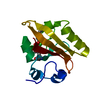
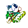
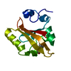
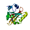
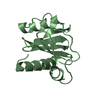
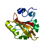
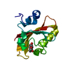
| Title | Photoactivated chromophore conformation in Photoactive Yellow Protein (E46Q mutant) from 10 microseconds to 3 milliseconds | ||||||
 Components Components | Photoactive yellow protein | ||||||
 Keywords Keywords | PHOTORECEPTOR / room temperature / time-resolved | ||||||
| Function / homology |  Function and homology informationphotoreceptor activity / phototransduction / regulation of DNA-templated transcription / identical protein binding Function and homology informationphotoreceptor activity / phototransduction / regulation of DNA-templated transcription / identical protein bindingSimilarity search - Function | ||||||
| Biological species |  Halorhodospira halophila (bacteria) Halorhodospira halophila (bacteria) | ||||||
| Method | X-RAY DIFFRACTION / SYNCHROTRON / MOLECULAR REPLACEMENT / Resolution: 1.6 Å | ||||||
 Authors Authors | Anderson, S. / Srajer, V. / Pahl, R. / Rajagopal, S. / Schotte, F. / Anfinrud, P. / Wulff, M. / Moffat, K. | ||||||
 Citation Citation | Journal: Structure / Year: 2004 Title: Chromophore conformation and the evolution of tertiary structural changes in photoactive yellow protein Authors: Anderson, S. / Srajer, V. / Pahl, R. / Rajagopal, S. / Schotte, F. / Anfinrud, P. / Wulff, M. / Moffat, K. | ||||||
| History |
|
- Structure visualization
| Structure viewer | Molecule: MolmilJmol/JSmol |
|---|
- Downloads & links
-Download
| PDBx/mmCIF format | 1s1y.cif.gz | 41.4 KB | Display | PDBx/mmCIF format |
|---|---|---|---|---|
| PDB format | pdb1s1y.ent.gz | 32.1 KB | Display | PDB format |
| PDBx/mmJSON format | 1s1y.json.gz | Tree view | PDBx/mmJSON format | |
| Others |  Other downloads Other downloads |
-Validation report
| Arichive directory | https://data.pdbj.org/pub/pdb/validation_reports/s1/1s1yftp://data.pdbj.org/pub/pdb/validation_reports/s1/1s1y | HTTPS FTP |
|---|
-Related structure data


-Links
 PDBj
PDBj


- Assembly
| Deposited unit | 
| ||||||||
|---|---|---|---|---|---|---|---|---|---|
| 1 |
| ||||||||
| Unit cell |
|
-Components
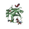
| #1: Protein | / PYP Mass: 13887.591 Da / Num. of mol.: 1 / Mutation: E46Q Source method: isolated from a genetically manipulated source Source: (gene. exp.) Halorhodospira halophila (bacteria) / Gene: PYP / Production host: Escherichia coli (E. coli) / References: UniProt: P16113 |
|---|---|
| #2: Chemical | ChemComp-HC4 / P-Coumaric acid  Mass: 164.158 Da / Num. of mol.: 1 / Source method: obtained synthetically / Formula: C9H8O3 Mass: 164.158 Da / Num. of mol.: 1 / Source method: obtained synthetically / Formula: C9H8O3 |
| #3: Water | ChemComp-HOH / Water Mass: 18.015 Da / Num. of mol.: 111 / Source method: isolated from a natural source / Formula: H2O Mass: 18.015 Da / Num. of mol.: 111 / Source method: isolated from a natural source / Formula: H2O |
-Experimental details
-Experiment
| Experiment | Method: X-RAY DIFFRACTION / Number of used crystals: 15 |
|---|
- Sample preparation
| Crystal | Density Matthews: 1.91 Å3/Da / Density % sol: 35.52 % |
|---|---|
| Crystal grow | Temperature: 293 K / Method: vapor diffusion, hanging drop / pH: 7 Details: ammonium sulfate, sodium phosphate, pH 7.0, VAPOR DIFFUSION, HANGING DROP, temperature 293K |
-Data collection
| Diffraction |
| |||||||||||||||||||||||||
|---|---|---|---|---|---|---|---|---|---|---|---|---|---|---|---|---|---|---|---|---|---|---|---|---|---|---|
| Diffraction source |
| |||||||||||||||||||||||||
| Detector |
| |||||||||||||||||||||||||
| Radiation |
| |||||||||||||||||||||||||
| Radiation wavelength | Relative weight: 1 | |||||||||||||||||||||||||
| Reflection | Resolution: 1.6→100 Å / Num. obs: 13371 / % possible obs: 95.8 % / Observed criterion σ(F): 2 / Observed criterion σ(I): 2 | |||||||||||||||||||||||||
| Reflection shell | Resolution: 1.6→1.67 Å / % possible all: 83.8 |


- Processing
| Software |
| |||||||||||||||||||||||||
|---|---|---|---|---|---|---|---|---|---|---|---|---|---|---|---|---|---|---|---|---|---|---|---|---|---|---|
| Refinement | Method to determine structure: MOLECULAR REPLACEMENT / Resolution: 1.6→100 Å / Cross valid method: THROUGHOUT / σ(F): 4 / Stereochemistry target values: Engh & Huber / Details: difference refinement
| |||||||||||||||||||||||||
| Refinement step | Cycle: LAST / Resolution: 1.6→100 Å
|
