 Movie
Movie Controller

[English] 日本語
 Yorodumi
- PDB-1s0h: Structure determination of haemoglobin from Donkey(equus asinus) ... -
+ Open data

- Basic information
| Entry | Database: PDB / ID: 1s0h | ||||||
|---|---|---|---|---|---|---|---|
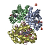
| Title | Structure determination of haemoglobin from Donkey(equus asinus) at 3.0 Angstrom resolution | ||||||
 Components Components |
| ||||||
 Keywords Keywords | OXYGEN STORAGE/TRANSPORT /  alpha helix / dimer / OXYGEN STORAGE-TRANSPORT COMPLEX alpha helix / dimer / OXYGEN STORAGE-TRANSPORT COMPLEX | ||||||
| Function / homology |  Function and homology informationhemoglobin complex / oxygen carrier activity / oxygen binding / iron ion binding / heme binding / metal ion binding Function and homology informationhemoglobin complex / oxygen carrier activity / oxygen binding / iron ion binding / heme binding / metal ion bindingSimilarity search - Function | ||||||
| Biological species |  Equus asinus (ass) Equus asinus (ass) | ||||||
| Method | X-RAY DIFFRACTION / MOLECULAR REPLACEMENT / Resolution: 3 Å | ||||||
 Authors Authors | Balasundaresan, D. / Ponnuswamy, M.N. / Saraboji, K. | ||||||
 Citation Citation | Journal: Biochimie / Year: 2006 Title: Crystal structure of haemoglobin from donkey (Equus asinus) at 3A resolution Authors: Balasundaresan, D. / Saraboji, K. / Ponnuswamy, M.N. | ||||||
| History |
|
- Structure visualization
| Structure viewer | Molecule: MolmilJmol/JSmol |
|---|
- Downloads & links
-Download
| PDBx/mmCIF format | 1s0h.cif.gz | 70.7 KB | Display | PDBx/mmCIF format |
|---|---|---|---|---|
| PDB format | pdb1s0h.ent.gz | 52.5 KB | Display | PDB format |
| PDBx/mmJSON format | 1s0h.json.gz | Tree view | PDBx/mmJSON format | |
| Others |  Other downloads Other downloads |
-Validation report
| Arichive directory | https://data.pdbj.org/pub/pdb/validation_reports/s0/1s0hftp://data.pdbj.org/pub/pdb/validation_reports/s0/1s0h | HTTPS FTP |
|---|
-Related structure data
| Related structure data |  2mhbS S: Starting model for refinement |
|---|---|
| Similar structure data |


-Links
 PDBj
PDBj


- Assembly
| Deposited unit | 
| ||||||||
|---|---|---|---|---|---|---|---|---|---|
| 1 | 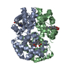
| ||||||||
| Unit cell |
| ||||||||
| Details | TO GENERATE COORDINATES FOR THE ALPHA-2 AND BETA-2 CHAINS FROM THE ALPHA-1 AND BETA-1 COORDINATES GIVEN HERE, APPLY THE OPERATION: -x,y,-z |
-Components
| #1: Protein | Mass: 15098.237 Da / Num. of mol.: 1 / Source method: isolated from a natural source / Source: (natural) Equus asinus (ass) / Cell: Erythrocytes / References: UniProt: P01959 |
|---|---|
| #2: Protein | Mass: 16032.274 Da / Num. of mol.: 1 / Source method: isolated from a natural source / Source: (natural) Equus asinus (ass) / Cell: Erythrocytes / References: UniProt: D1MPT0*PLUS |
| #3: Chemical | Heme B  Mass: 616.487 Da / Num. of mol.: 2 / Source method: obtained synthetically / Formula: C34H32FeN4O4 Mass: 616.487 Da / Num. of mol.: 2 / Source method: obtained synthetically / Formula: C34H32FeN4O4 |
-Experimental details
-Experiment
| Experiment | Method: X-RAY DIFFRACTION / Number of used crystals: 2 |
|---|
- Sample preparation
| Crystal | Density Matthews: 2.67 Å3/Da / Density % sol: 53.49 % |
|---|---|
| Crystal grow | Temperature: 293 K / Method: vapor diffusion, hanging drop / pH: 6.8 Details: 2.8M Na/K phosphate buffer, pH 6.8, VAPOR DIFFUSION, HANGING DROP, temperature 293K |
-Data collection
| Diffraction | Mean temperature: 298 K |
|---|---|
| Diffraction source | Source: ROTATING ANODE / Type: RIGAKU RU200 / Wavelength: 1.5418 Å |
| Detector | Type: MARRESEARCH / Detector: IMAGE PLATE / Date: Oct 8, 2003 / Details: Monochromator |
| Radiation | Monochromator: M / Protocol: SINGLE WAVELENGTH / Monochromatic (M) / Laue (L): M / Scattering type: x-ray |
| Radiation wavelength | Wavelength: 1.5418 Å / Relative weight: 1 |
| Reflection | Resolution: 3.002→30 Å / Num. all: 5406 / Num. obs: 5387 / % possible obs: 79.01 % / Observed criterion σ(F): 2 / Observed criterion σ(I): 2 / Redundancy: 4.1 % / Rmerge(I) obs: 0.161 / Net I/σ(I): 4.3 |
| Reflection shell | Resolution: 3.002→3.079 Å / Rmerge(I) obs: 0.43 / Num. unique all: 584 / % possible all: 34.3 |
- Processing
| Software |
| ||||||||||||||||||||||||||||||||||||||||||||||||||||||||||||||||||||||||||||||||||||||||||||||||||||
|---|---|---|---|---|---|---|---|---|---|---|---|---|---|---|---|---|---|---|---|---|---|---|---|---|---|---|---|---|---|---|---|---|---|---|---|---|---|---|---|---|---|---|---|---|---|---|---|---|---|---|---|---|---|---|---|---|---|---|---|---|---|---|---|---|---|---|---|---|---|---|---|---|---|---|---|---|---|---|---|---|---|---|---|---|---|---|---|---|---|---|---|---|---|---|---|---|---|---|---|---|---|
| Refinement | Method to determine structure: MOLECULAR REPLACEMENT Starting model: Horse Methaemoglobin(PDB ID:2MHB) Resolution: 3→21.18 Å / Cor.coef. Fo:Fc: 0.935 / Cor.coef. Fo:Fc free: 0.828 / SU B: 32.603 / SU ML: 0.545 / Cross valid method: THROUGHOUT / σ(F): 2 / ESU R Free: 0.628 / Stereochemistry target values: MAXIMUM LIKELIHOOD / Details: HYDROGENS HAVE BEEN ADDED IN THE RIDING POSITIONS
| ||||||||||||||||||||||||||||||||||||||||||||||||||||||||||||||||||||||||||||||||||||||||||||||||||||
| Solvent computation | Ion probe radii: 0.8 Å / Shrinkage radii: 0.8 Å / VDW probe radii: 1.4 Å / Solvent model: BABINET MODEL WITH MASK | ||||||||||||||||||||||||||||||||||||||||||||||||||||||||||||||||||||||||||||||||||||||||||||||||||||
| Displacement parameters | Biso mean: 33.322 Å2
| ||||||||||||||||||||||||||||||||||||||||||||||||||||||||||||||||||||||||||||||||||||||||||||||||||||
| Refinement step | Cycle: LAST / Resolution: 3→21.18 Å
| ||||||||||||||||||||||||||||||||||||||||||||||||||||||||||||||||||||||||||||||||||||||||||||||||||||
| Refine LS restraints |
| ||||||||||||||||||||||||||||||||||||||||||||||||||||||||||||||||||||||||||||||||||||||||||||||||||||
| LS refinement shell | Resolution: 3.002→3.079 Å / Total num. of bins used: 20
|
