 Movie
Movie Controller


+ Open data

- Basic information
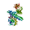
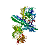
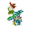
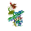
| Entry | Database: PDB / ID: 1s0d | ||||||
|---|---|---|---|---|---|---|---|
| Title | Crystal structure of botulinum neurotoxin type B at pH 5.5 | ||||||
 Components Components | Botulinum neurotoxin type B | ||||||
 Keywords Keywords |  Toxin / hydrolase / botulinum / neurotoxin / pH / metals Toxin / hydrolase / botulinum / neurotoxin / pH / metals | ||||||
| Function / homology |  Function and homology information Function and homology informationToxicity of botulinum toxin type B (botB) / bontoxilysin / host cell presynaptic membrane / host cell cytoplasmic vesicle / host cell cytosol / protein transmembrane transporter activity / metalloendopeptidase activity / toxin activity / lipid binding / host cell plasma membrane ...Toxicity of botulinum toxin type B (botB) / bontoxilysin / host cell presynaptic membrane / host cell cytoplasmic vesicle / host cell cytosol / protein transmembrane transporter activity / metalloendopeptidase activity / toxin activity / lipid binding / host cell plasma membrane / proteolysis / zinc ion binding / extracellular region / membraneSimilarity search - Function | ||||||
| Biological species |   Clostridium botulinum (bacteria) Clostridium botulinum (bacteria) | ||||||
| Method | X-RAY DIFFRACTION / SYNCHROTRON / MOLECULAR REPLACEMENT / Resolution: 2.2 Å | ||||||
 Authors Authors | Eswaramoorthy, S. / Kumaran, D. / Keller, J. / Swaminathan, S. | ||||||
 Citation Citation | Journal: Biochemistry / Year: 2004 Title: Role of metals in the biological activity of Clostridium botulinum neurotoxins Authors: Eswaramoorthy, S. / Kumaran, D. / Keller, J. / Swaminathan, S. | ||||||
| History |
|
- Structure visualization
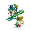
| Structure viewer | Molecule: MolmilJmol/JSmol |
|---|
- Downloads & links
-Download
| PDBx/mmCIF format | 1s0d.cif.gz | 283.8 KB | Display | PDBx/mmCIF format |
|---|---|---|---|---|
| PDB format | pdb1s0d.ent.gz | 225.3 KB | Display | PDB format |
| PDBx/mmJSON format | 1s0d.json.gz | Tree view | PDBx/mmJSON format | |
| Others |  Other downloads Other downloads |
-Validation report
| Arichive directory | https://data.pdbj.org/pub/pdb/validation_reports/s0/1s0dftp://data.pdbj.org/pub/pdb/validation_reports/s0/1s0d | HTTPS FTP |
|---|
-Related structure data
| Related structure data |  1s0bC  1s0cC  1s0eC  1s0fC  1s0gC  1epwS S: Starting model for refinement C: citing same article ( |
|---|---|
| Similar structure data |


-Links
 PDBj
PDBj- Assembly
| Deposited unit | 
| ||||||||
|---|---|---|---|---|---|---|---|---|---|
| 1 |
| ||||||||
| Unit cell |
|
-Components
| #1: Protein | Mass: 150833.375 Da / Num. of mol.: 1 / Source method: isolated from a natural source / Source: (natural) Clostridium botulinum (bacteria) / Strain: type B / References: UniProt: P10844, bontoxilysin | ||
|---|---|---|---|
| #2: Chemical | ChemComp-ZN /   Mass: 65.409 Da / Num. of mol.: 1 / Source method: obtained synthetically / Formula: Zn Mass: 65.409 Da / Num. of mol.: 1 / Source method: obtained synthetically / Formula: Zn | ||
| #3: Chemical |   Mass: 40.078 Da / Num. of mol.: 2 / Source method: obtained synthetically / Formula: Ca Mass: 40.078 Da / Num. of mol.: 2 / Source method: obtained synthetically / Formula: Ca#4: Water | ChemComp-HOH / | Water Mass: 18.015 Da / Num. of mol.: 547 / Source method: isolated from a natural source / Formula: H2O Mass: 18.015 Da / Num. of mol.: 547 / Source method: isolated from a natural source / Formula: H2O |
-Experimental details
-Experiment
| Experiment | Method: X-RAY DIFFRACTION / Number of used crystals: 1 |
|---|
- Sample preparation
| Crystal | Density Matthews: 2.78 Å3/Da / Density % sol: 55.79 % | ||||||||||||||||||||||||
|---|---|---|---|---|---|---|---|---|---|---|---|---|---|---|---|---|---|---|---|---|---|---|---|---|---|
| Crystal grow | Temperature: 298 K / Method: vapor diffusion, sitting drop / pH: 5.5 Details: PEG 4000, MES, pH 5.5, VAPOR DIFFUSION, SITTING DROP, temperature 298K | ||||||||||||||||||||||||
| Crystal grow | *PLUS pH: 6 / Method: vapor diffusionDetails: Swaminathan, S., (2000) Acta Crystallogr., Sect.D, 56, 1024. | ||||||||||||||||||||||||
| Components of the solutions | *PLUS
|
-Data collection
| Diffraction | Mean temperature: 100 K |
|---|---|
| Diffraction source | Source: SYNCHROTRON / Site: NSLS  / Beamline: X12C / Wavelength: 1 Å / Beamline: X12C / Wavelength: 1 Å |
| Detector | Type: BRANDEIS - B1.2 / Detector: CCD |
| Radiation | Protocol: SINGLE WAVELENGTH / Monochromatic (M) / Laue (L): M / Scattering type: x-ray |
| Radiation wavelength | Wavelength: 1 Å / Relative weight: 1 |
| Reflection | Resolution: 2.2→50 Å / Num. all: 78587 / Num. obs: 78587 / % possible obs: 94.3 % / Observed criterion σ(F): 0 / Observed criterion σ(I): 0 / Redundancy: 3.3 % / Rmerge(I) obs: 0.067 / Net I/σ(I): 8.1 |
| Reflection shell | Resolution: 2.2→2.28 Å / Rmerge(I) obs: 0.397 / Num. unique all: 6230 / % possible all: 74.8 |
| Reflection | *PLUS Num. measured all: 275095 |
| Reflection shell | *PLUS % possible obs: 74.8 % |
- Processing
| Software |
| |||||||||||||||||||||||||
|---|---|---|---|---|---|---|---|---|---|---|---|---|---|---|---|---|---|---|---|---|---|---|---|---|---|---|
| Refinement | Method to determine structure: MOLECULAR REPLACEMENT Starting model: PDB ENTRY 1EPW Resolution: 2.2→50 Å / Cross valid method: THROUGHOUT / σ(F): 0 / Stereochemistry target values: Engh & Huber
| |||||||||||||||||||||||||
| Refinement step | Cycle: LAST / Resolution: 2.2→50 Å
| |||||||||||||||||||||||||
| Refine LS restraints |
| |||||||||||||||||||||||||
| Refinement | *PLUS % reflection Rfree: 2 % / Rfactor Rfree: 0.273 / Rfactor Rwork: 0.223 | |||||||||||||||||||||||||
| Solvent computation | *PLUS | |||||||||||||||||||||||||
| Displacement parameters | *PLUS | |||||||||||||||||||||||||
| Refine LS restraints | *PLUS
|
