 Movie
Movie Controller

[English] 日本語
 Yorodumi
- PDB-1ryg: Three dimensional solution structure of the R29A MUTANT of sodium... -
+ Open data

- Basic information
| Entry | Database: PDB / ID: 1ryg | ||||||
|---|---|---|---|---|---|---|---|
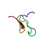
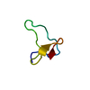
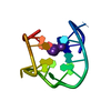
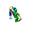
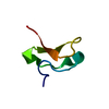
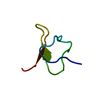
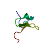
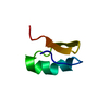
| Title | Three dimensional solution structure of the R29A MUTANT of sodium channels inhibitor HAINANTOXIN-IV by 2D 1H-NMR | ||||||
 Components Components | Hainantoxin-IV | ||||||
 Keywords Keywords | TOXIN / Neurotoxin / Inhibitor cystine knot motif | ||||||
| Function / homology |  Function and homology information Function and homology informationhost cell presynaptic membrane / ion channel inhibitor activity / sodium channel regulator activity / toxin activity / extracellular regionSimilarity search - Function | ||||||
| Method | SOLUTION NMR / simulated annealing | ||||||
 Authors Authors | Li, D. / Lu, S. / Gu, X. / Liang, S. | ||||||
 Citation Citation | Journal: J.Biol.Chem. / Year: 2004 Title: Structure--activity relationships of hainantoxin-IV and structure determination of active and inactive sodium channel blockers Authors: Li, D. / Xiao, Y. / Xu, X. / Xiong, X. / Lu, S. / Liu, Z. / Zhu, Q. / Wang, M. / Gu, X. / Liang, S. #1: Journal: Acta Biochim.Biophys.Sinica / Year: 2002Title: Synthesis and Oxidative Refolding of Hainantoxin-Iv Authors: Liu, Z.H. / Chen, P. / Liang, S.P. | ||||||
| History |
|
- Structure visualization
| Structure viewer | Molecule: MolmilJmol/JSmol |
|---|
- Downloads & links
-Download
| PDBx/mmCIF format | 1ryg.cif.gz | 204.7 KB | Display | PDBx/mmCIF format |
|---|---|---|---|---|
| PDB format | pdb1ryg.ent.gz | 178 KB | Display | PDB format |
| PDBx/mmJSON format | 1ryg.json.gz | Tree view | PDBx/mmJSON format | |
| Others |  Other downloads Other downloads |
-Validation report
| Arichive directory | https://data.pdbj.org/pub/pdb/validation_reports/ry/1rygftp://data.pdbj.org/pub/pdb/validation_reports/ry/1ryg | HTTPS FTP |
|---|
-Related structure data


-Links
 PDBj
PDBj- Assembly
| Deposited unit | 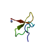
| |||||||||
|---|---|---|---|---|---|---|---|---|---|---|
| 1 |
| |||||||||
| NMR ensembles |
|
-Components
| #1: Protein/peptide | / HnTx-IV Mass: 3913.513 Da / Num. of mol.: 1 / Mutation: R29A / Source method: obtained synthetically / Details: synthesized using standard fmoc chemistry. / References: UniProt: P83471, UniProt: D2Y232*PLUS |
|---|
-Experimental details
-Experiment
| Experiment | Method: SOLUTION NMR | ||||||||||||||||
|---|---|---|---|---|---|---|---|---|---|---|---|---|---|---|---|---|---|
| NMR experiment |
| ||||||||||||||||
| NMR details | Text: this structure was determined using standard 2D homonuclear techniques. |
- Sample preparation
| Details | Contents: 5.0mM Solvent system: 20mM deuterium acetic acid BU, 90% H2O, 10% D2O |
|---|---|
| Sample conditions | Ionic strength: 20 / pH: 4.00 / Pressure: 1 atm / Temperature: 295 K |
-NMR measurement
| Radiation | Protocol: SINGLE WAVELENGTH / Monochromatic (M) / Laue (L): M |
|---|---|
| Radiation wavelength | Relative weight: 1 |
| NMR spectrometer | Type: Bruker DRX / Manufacturer: Bruker / Model: DRX / Field strength: 500 MHz |
- Processing
| NMR software |
| |||||||||
|---|---|---|---|---|---|---|---|---|---|---|
| Refinement | Method: simulated annealing / Software ordinal: 1 Details: the structures are based on 366 NOE- derived distance constraints, 15 dihedral angle restraints, 9 fake distance restraints from disulfide bonds and 12 hydrogen- bond constrains. | |||||||||
| NMR representative | Selection criteria: lowest energy | |||||||||
| NMR ensemble | Conformer selection criteria: structures with the lowest energy Conformers calculated total number: 50 / Conformers submitted total number: 20 |
