 Movie
Movie Controller

[English] 日本語
 Yorodumi
- PDB-1ry1: Structure of the signal recognition particle interacting with the... -
+ Open data

- Basic information
| Entry | Database: PDB / ID: 1ry1 | ||||||
|---|---|---|---|---|---|---|---|
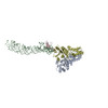
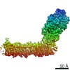
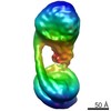
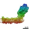
| Title | Structure of the signal recognition particle interacting with the elongation-arrested ribosome | ||||||
 Components Components |
| ||||||
 Keywords Keywords | TRANSLATION / signal recognition particle / RNA binding | ||||||
| Function / homology |  Function and homology information Function and homology informationsignal recognition particle receptor complex / SRP-dependent cotranslational protein targeting to membrane / SRP-dependent cotranslational protein targeting to membrane, signal sequence recognition / signal recognition particle, endoplasmic reticulum targeting / endoplasmic reticulum signal peptide binding / granulocyte differentiation / absorption of visible light / signal recognition particle binding / G protein-coupled opsin signaling pathway / signal recognition particle ...signal recognition particle receptor complex / SRP-dependent cotranslational protein targeting to membrane / SRP-dependent cotranslational protein targeting to membrane, signal sequence recognition / signal recognition particle, endoplasmic reticulum targeting / endoplasmic reticulum signal peptide binding / granulocyte differentiation / absorption of visible light / signal recognition particle binding / G protein-coupled opsin signaling pathway / signal recognition particle / photoreceptor inner segment membrane / 11-cis retinal binding / cotranslational protein targeting to membrane / G protein-coupled photoreceptor activity / signal-recognition-particle GTPase / negative regulation of translational elongation / protein targeting to ER / 7S RNA binding / exocrine pancreas development / SRP-dependent cotranslational protein targeting to membrane, translocation / SRP-dependent cotranslational protein targeting to membrane / photoreceptor outer segment membrane / SRP-dependent cotranslational protein targeting to membrane / ribonucleoprotein complex binding / visual perception / neutrophil chemotaxis / photoreceptor disc membrane / secretory granule lumen / ficolin-1-rich granule lumen / nuclear body / nuclear speck / GTPase activity / Neutrophil degranulation / nucleolus / GTP binding / endoplasmic reticulum / ATP hydrolysis activity / RNA binding / extracellular region / membrane / nucleus / metal ion binding / plasma membrane / cytoplasm / cytosol Similarity search - Function | ||||||
| Biological species |   | ||||||
| Method | ELECTRON MICROSCOPY / single particle reconstruction / cryo EM / Resolution: 12 Å | ||||||
 Authors Authors | Halic, M. / Becker, T. / Pool, M.R. / Spahn, C.M. / Grassucci, R.A. / Frank, J. / Beckmann, R. | ||||||
 Citation Citation | Journal: Nature / Year: 2004 Title: Structure of the signal recognition particle interacting with the elongation-arrested ribosome. Authors: Mario Halic / Thomas Becker / Martin R Pool / Christian M T Spahn / Robert A Grassucci / Joachim Frank / Roland Beckmann /  Abstract: Cotranslational translocation of proteins across or into membranes is a vital process in all kingdoms of life. It requires that the translating ribosome be targeted to the membrane by the signal ...Cotranslational translocation of proteins across or into membranes is a vital process in all kingdoms of life. It requires that the translating ribosome be targeted to the membrane by the signal recognition particle (SRP), an evolutionarily conserved ribonucleoprotein particle. SRP recognizes signal sequences of nascent protein chains emerging from the ribosome. Subsequent binding of SRP leads to a pause in peptide elongation and to the ribosome docking to the membrane-bound SRP receptor. Here we present the structure of a targeting complex consisting of mammalian SRP bound to an active 80S ribosome carrying a signal sequence. This structure, solved to 12 A by cryo-electron microscopy, enables us to generate a molecular model of SRP in its functional conformation. The model shows how the S domain of SRP contacts the large ribosomal subunit at the nascent chain exit site to bind the signal sequence, and that the Alu domain reaches into the elongation-factor-binding site of the ribosome, explaining its elongation arrest activity. | ||||||
| History |
|
- Structure visualization
| Movie |
Movie viewer |
|---|---|
| Structure viewer | Molecule: MolmilJmol/JSmol |
- Downloads & links
-Download
| PDBx/mmCIF format | 1ry1.cif.gz | 310 KB | Display | PDBx/mmCIF format |
|---|---|---|---|---|
| PDB format | pdb1ry1.ent.gz | 231.9 KB | Display | PDB format |
| PDBx/mmJSON format | 1ry1.json.gz | Tree view | PDBx/mmJSON format | |
| Others |  Other downloads Other downloads |
-Validation report
| Summary document | 1ry1_validation.pdf.gz | 927.2 KB | Display | wwPDB validaton report |
|---|---|---|---|---|
| Full document | 1ry1_full_validation.pdf.gz | 1 MB | Display | |
| Data in XML | 1ry1_validation.xml.gz | 42.3 KB | Display | |
| Data in CIF | 1ry1_validation.cif.gz | 64.1 KB | Display | |
| Arichive directory | https://data.pdbj.org/pub/pdb/validation_reports/ry/1ry1ftp://data.pdbj.org/pub/pdb/validation_reports/ry/1ry1 | HTTPS FTP |
-Related structure data
| Related structure data | 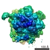 1063MC M: map data used to model this data C: citing same article ( |
|---|---|
| Similar structure data |
-Links
 PDBj
PDBj


- Assembly
| Deposited unit | 
|
|---|---|
| 1 |
|
-Components
-RNA chain , 8 types, 8 molecules EAMNOPQR
| #1: RNA chain | Mass: 16277.622 Da / Num. of mol.: 1 / Source method: isolated from a natural source / Source: (natural) |
|---|---|
| #2: RNA chain | Mass: 41566.715 Da / Num. of mol.: 1 / Source method: isolated from a natural source / Source: (natural) |
| #3: RNA chain | Mass: 8784.278 Da / Num. of mol.: 1 / Source method: isolated from a natural source / Source: (natural) |
| #4: RNA chain | Mass: 9807.837 Da / Num. of mol.: 1 / Source method: isolated from a natural source / Source: (natural) |
| #5: RNA chain | Mass: 7695.542 Da / Num. of mol.: 1 / Source method: isolated from a natural source / Source: (natural) |
| #6: RNA chain | Mass: 6325.847 Da / Num. of mol.: 1 / Source method: isolated from a natural source / Source: (natural) |
| #7: RNA chain | Mass: 3844.320 Da / Num. of mol.: 1 / Source method: isolated from a natural source / Source: (natural) |
| #8: RNA chain | Mass: 3804.296 Da / Num. of mol.: 1 / Source method: isolated from a natural source / Source: (natural) |
-Protein , 5 types, 5 molecules CDBUW
| #9: Protein | Mass: 9996.567 Da / Num. of mol.: 1 / Source method: isolated from a natural source / Source: (natural) |
|---|---|
| #10: Protein | Mass: 12114.235 Da / Num. of mol.: 1 / Source method: isolated from a natural source / Source: (natural) |
| #11: Protein | Mass: 12561.688 Da / Num. of mol.: 1 / Source method: isolated from a natural source / Source: (natural) |
| #12: Protein | Mass: 32472.508 Da / Num. of mol.: 1 / Source method: isolated from a natural source / Source: (natural) |
| #13: Protein | Mass: 12473.440 Da / Num. of mol.: 1 / Source method: isolated from a natural source / Source: (natural) |
-Protein/peptide , 1 types, 1 molecules S
| #14: Protein/peptide | Mass: 2121.542 Da / Num. of mol.: 1 / Source method: obtained synthetically / Source: (synth.) |
|---|
-Details
| Sequence details | This structure has been modeled using polymer sequences from other organisms, including Homo ...This structure has been modeled using polymer sequences from other organisms, including Homo sapiens (chains C,D,E), Thermus aquaticus (chain U), Mus musculus (chain W), and Tursiops truncatus (chain S). |
|---|
-Experimental details
-Experiment
| Experiment | Method: ELECTRON MICROSCOPY |
|---|---|
| EM experiment | Aggregation state: PARTICLE / 3D reconstruction method: single particle reconstruction |
- Sample preparation
| Component |
| ||||||||||||||||||||||||
|---|---|---|---|---|---|---|---|---|---|---|---|---|---|---|---|---|---|---|---|---|---|---|---|---|---|
| Source (natural) |
| ||||||||||||||||||||||||
| Buffer solution | pH: 7.4 | ||||||||||||||||||||||||
| Specimen | Embedding applied: NO / Shadowing applied: NO / Staining applied: NO / Vitrification applied: YES | ||||||||||||||||||||||||
| Specimen support | Details: HOLEY CARBON | ||||||||||||||||||||||||
| Vitrification | Cryogen name: ETHANE / Details: PLUNGED INTO ETHANE |
- Electron microscopy imaging
| Experimental equipment |  Model: Tecnai F20 / Image courtesy: FEI Company |
|---|---|
| Microscopy | Model: FEI TECNAI F20 / Date: Jan 1, 2000 Details: SAMPLES WERE MAINTAINED AT LIQUID NITROGEN TEMPERATURES IN THE ELECTRON MICROSCOPE. |
| Electron gun | Electron source:  FIELD EMISSION GUN / Accelerating voltage: 160 kV / Illumination mode: FLOOD BEAM FIELD EMISSION GUN / Accelerating voltage: 160 kV / Illumination mode: FLOOD BEAM |
| Electron lens | Mode: BRIGHT FIELD / Nominal magnification: 51000 X / Calibrated magnification: 52000 X / Nominal defocus max: 45000 nm / Nominal defocus min: 10000 nm / Cs: 2 mm |
| Specimen holder | Temperature: 95 K / Tilt angle max: 0 ° / Tilt angle min: 0 ° |
| Image recording | Electron dose: 10 e/Å2 / Film or detector model: KODAK SO-163 FILM |
| Image scans | Num. digital images: 75 |
- Processing
| EM software |
| ||||||||||||
|---|---|---|---|---|---|---|---|---|---|---|---|---|---|
| Symmetry | Point symmetry: C1 (asymmetric) | ||||||||||||
| 3D reconstruction | Resolution: 12 Å / Resolution method: FSC 0.5 CUT-OFF Details: The chains M, N, O, P, Q and R are fragments of a double helical strand of RNA. The author maintains that some of the residues could not be modeled correctly due to limited resolution in this region. Symmetry type: POINT | ||||||||||||
| Refinement | Highest resolution: 12 Å | ||||||||||||
| Refinement step | Cycle: LAST / Highest resolution: 12 Å
|
