 Movie
Movie Controller

[English] 日本語
 Yorodumi
- PDB-1rt8: CRYSTAL STRUCTURE OF THE ACTIN-CROSSLINKING CORE OF SCHIZOSACCHAR... -
+ Open data

- Basic information
| Entry | Database: PDB / ID: 1rt8 | ||||||
|---|---|---|---|---|---|---|---|
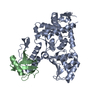
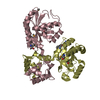
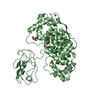
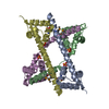
| Title | CRYSTAL STRUCTURE OF THE ACTIN-CROSSLINKING CORE OF SCHIZOSACCHAROMYCES POMBE FIMBRIN | ||||||
 Components Components | fimbrin | ||||||
 Keywords Keywords |  STRUCTURAL PROTEIN / Filamentous Actin Binding Domain (ABD) / Calponin Homology / Actin-Crosslinking STRUCTURAL PROTEIN / Filamentous Actin Binding Domain (ABD) / Calponin Homology / Actin-Crosslinking | ||||||
| Function / homology |  Function and homology information Function and homology informationactomyosin contractile ring organization / actin body / formin-nucleated actin cable organization / mitotic actomyosin contractile ring, distal actin filament layer / positive regulation of actin filament severing / negative regulation of actin filament binding / actin cortical patch organization / medial cortex / actin cortical patch localization / actin filament network formation ...actomyosin contractile ring organization / actin body / formin-nucleated actin cable organization / mitotic actomyosin contractile ring, distal actin filament layer / positive regulation of actin filament severing / negative regulation of actin filament binding / actin cortical patch organization / medial cortex / actin cortical patch localization / actin filament network formation / negative regulation of Arp2/3 complex-mediated actin nucleation / actin cortical patch / cell division site / actin filament bundle / actin filament bundle assembly / actin filament / endocytosis / actin filament binding / calcium ion binding / cytoplasmSimilarity search - Function | ||||||
| Biological species |  Schizosaccharomyces pombe (fission yeast) Schizosaccharomyces pombe (fission yeast) | ||||||
| Method | X-RAY DIFFRACTION / SYNCHROTRON / AB INITIO PHASING / Resolution: 2 Å | ||||||
 Authors Authors | Klein, M.G. / Shi, W. / Ramagopal, U. / Tseng, Y. / Wirtz, D. / Kovar, D.R. / Staiger, C.J. / Almo, S.C. | ||||||
 Citation Citation | Journal: Structure / Year: 2004 Title: Structure of the actin crosslinking core of fimbrin. Authors: Klein, M.G. / Shi, W. / Ramagopal, U. / Tseng, Y. / Wirtz, D. / Kovar, D.R. / Staiger, C.J. / Almo, S.C. #1: Journal: Mol.Cell.Biol. / Year: 2001Title: Interactions among a fibrin, a capping protein, and an actin-depolymerizing factor in organization of the fission yeast actin cytoskeleton Authors: Nakano, K. / Satoh, K. / Morimatsu, A. / Ohnuma, M. / Mabuchi, I. | ||||||
| History |
|
- Structure visualization
| Structure viewer | Molecule: MolmilJmol/JSmol |
|---|
- Downloads & links
-Download
| PDBx/mmCIF format | 1rt8.cif.gz | 106.1 KB | Display | PDBx/mmCIF format |
|---|---|---|---|---|
| PDB format | pdb1rt8.ent.gz | 80.4 KB | Display | PDB format |
| PDBx/mmJSON format | 1rt8.json.gz | Tree view | PDBx/mmJSON format | |
| Others |  Other downloads Other downloads |
-Validation report
| Arichive directory | https://data.pdbj.org/pub/pdb/validation_reports/rt/1rt8ftp://data.pdbj.org/pub/pdb/validation_reports/rt/1rt8 | HTTPS FTP |
|---|
-Related structure data
| Related structure data | 1pxySC S: Starting model for refinement C: citing same article ( |
|---|---|
| Similar structure data |


-Links
 PDBj
PDBj- Assembly
| Deposited unit | 
| ||||||||
|---|---|---|---|---|---|---|---|---|---|
| 1 |
| ||||||||
| Unit cell |
|
-Components
| #1: Protein | Mass: 57451.445 Da / Num. of mol.: 1 / Fragment: Actin-Crosslinking Core, amino acids 108-614 Source method: isolated from a genetically manipulated source Source: (gene. exp.) Schizosaccharomyces pombe (fission yeast)Plasmid: pGEX-4T-1 / Species (production host): Escherichia coli / Production host:  Escherichia coli BL21 (bacteria) / Strain (production host): BL21 / References: UniProt: O59945 Escherichia coli BL21 (bacteria) / Strain (production host): BL21 / References: UniProt: O59945 | ||
|---|---|---|---|
| #2: Chemical | ChemComp-SO4 / Sulfate  Mass: 96.063 Da / Num. of mol.: 5 / Source method: obtained synthetically / Formula: SO4 Mass: 96.063 Da / Num. of mol.: 5 / Source method: obtained synthetically / Formula: SO4#3: Water | ChemComp-HOH / | Water Mass: 18.015 Da / Num. of mol.: 104 / Source method: isolated from a natural source / Formula: H2O Mass: 18.015 Da / Num. of mol.: 104 / Source method: isolated from a natural source / Formula: H2O |
-Experimental details
-Experiment
| Experiment | Method: X-RAY DIFFRACTION / Number of used crystals: 2 |
|---|
- Sample preparation
| Crystal | Density Matthews: 2.69 Å3/Da / Density % sol: 54.24 % |
|---|---|
| Crystal grow | Temperature: 298 K / Method: vapor diffusion, hanging drop / pH: 5.7 Details: PEG 3300, MES, Lithium Sulfate, pH 5.7, VAPOR DIFFUSION, HANGING DROP, temperature 298K |
-Data collection
| Diffraction |
| |||||||||||||||
|---|---|---|---|---|---|---|---|---|---|---|---|---|---|---|---|---|
| Diffraction source |
| |||||||||||||||
| Detector |
| |||||||||||||||
| Radiation | Protocol: SINGLE WAVELENGTH / Monochromatic (M) / Laue (L): M / Scattering type: x-ray | |||||||||||||||
| Radiation wavelength |
| |||||||||||||||
| Reflection | Resolution: 2→30 Å / Num. all: 42732 / Num. obs: 37818 / % possible obs: 95.2 % / Observed criterion σ(F): 2 | |||||||||||||||
| Reflection shell | Resolution: 2→2.07 Å / % possible all: 65 |

- Processing
| Software |
| ||||||||||||||||||||
|---|---|---|---|---|---|---|---|---|---|---|---|---|---|---|---|---|---|---|---|---|---|
| Refinement | Method to determine structure: AB INITIO PHASING Starting model: PDB ENTRY 1PXY Resolution: 2→30 Å / σ(F): 2 / Stereochemistry target values: Engh & Huber
| ||||||||||||||||||||
| Displacement parameters | Biso mean: 40.7 Å2 | ||||||||||||||||||||
| Refinement step | Cycle: LAST / Resolution: 2→30 Å
|
