Resolution: 2.3→2.38 Å / Redundancy: 1.8 % / Rmerge(I) obs: 0.221 / Mean I/σ(I) obs: 4 / Num. unique all: 1067 / % possible all: 77
-
Processing
Software
Name
Version
Classification
REFMAC
5.1.24
refinement
HKL-2000
datareduction
SCALEPACK
datascaling
SOLVE
phasing
RESOLVE
phasing
Refinement
Method to determine structure: SIRAS / Resolution: 2.3→35.58 Å / Cor.coef. Fo:Fc: 0.95 / Cor.coef. Fo:Fc free: 0.932 / SU B: 5.543 / SU ML: 0.138 / Cross valid method: THROUGHOUT / σ(F): 0 / ESU R: 0.251 / ESU R Free: 0.208 Details: CNS was also used in refinement. Only the S for SO4 402 and 403 is present in the coordinates.
Rfactor
Num. reflection
% reflection
Selection details
Rfree
0.23637
1338
9.9 %
RANDOM
Rwork
0.19251
-
-
-
all
0.1971
13860
-
-
obs
0.19712
12210
99.21 %
-
Solvent computation
Ion probe radii: 0.8 Å / Shrinkage radii: 0.8 Å / VDW probe radii: 1.4 Å / Solvent model: BABINET MODEL WITH MASK
Displacement parameters
Biso mean: 47.441 Å2
Refine analyze
Free
Obs
Luzzati coordinate error
0.36 Å
0.3 Å
Luzzati d res low
-
5 Å
Luzzati sigma a
0.31 Å
0.24 Å
Refinement step
Cycle: LAST / Resolution: 2.3→35.58 Å
Protein
Nucleic acid
Ligand
Solvent
Total
Num. atoms
1561
0
13
58
1632
Refine LS restraints
Refine-ID
Type
Dev ideal
Dev ideal target
Number
X-RAY DIFFRACTION
r_bond_refined_d
0.036
0.021
1625
X-RAY DIFFRACTION
r_angle_refined_deg
2.382
1.967
2191
X-RAY DIFFRACTION
r_dihedral_angle_1_deg
6.671
5
195
X-RAY DIFFRACTION
r_chiral_restr
0.188
0.2
238
X-RAY DIFFRACTION
r_gen_planes_refined
0.013
0.02
1195
X-RAY DIFFRACTION
r_nbd_refined
0.26
0.2
706
X-RAY DIFFRACTION
r_xyhbond_nbd_refined
0.164
0.2
83
X-RAY DIFFRACTION
r_metal_ion_refined
0.166
0.2
3
X-RAY DIFFRACTION
r_symmetry_vdw_refined
0.733
0.2
49
X-RAY DIFFRACTION
r_symmetry_hbond_refined
0.312
0.2
9
X-RAY DIFFRACTION
r_mcbond_it
1.728
1.5
973
X-RAY DIFFRACTION
r_mcangle_it
3.151
2
1564
X-RAY DIFFRACTION
r_scbond_it
4.898
3
652
X-RAY DIFFRACTION
r_scangle_it
7.691
4.5
627
LS refinement shell
Resolution: 2.301→2.361 Å / Total num. of bins used: 20 /
Rfactor
Num. reflection
Rfree
0.327
80
Rwork
0.23
877
+
About Yorodumi
-
News
-
Feb 9, 2022. New format data for meta-information of EMDB entries
New format data for meta-information of EMDB entries
Version 3 of the EMDB header file is now the official format.
The previous official version 1.9 will be removed from the archive.
In the structure databanks used in Yorodumi, some data are registered as the other names, "COVID-19 virus" and "2019-nCoV". Here are the details of the virus and the list of structure data.
Jan 31, 2019. EMDB accession codes are about to change! (news from PDBe EMDB page)
EMDB accession codes are about to change! (news from PDBe EMDB page)
The allocation of 4 digits for EMDB accession codes will soon come to an end. Whilst these codes will remain in use, new EMDB accession codes will include an additional digit and will expand incrementally as the available range of codes is exhausted. The current 4-digit format prefixed with “EMD-” (i.e. EMD-XXXX) will advance to a 5-digit format (i.e. EMD-XXXXX), and so on. It is currently estimated that the 4-digit codes will be depleted around Spring 2019, at which point the 5-digit format will come into force.
The EM Navigator/Yorodumi systems omit the EMD- prefix.
Related info.:Q: What is EMD? / ID/Accession-code notation in Yorodumi/EM Navigator
Yorodumi is a browser for structure data from EMDB, PDB, SASBDB, etc.
This page is also the successor to EM Navigator detail page, and also detail information page/front-end page for Omokage search.
The word "yorodu" (or yorozu) is an old Japanese word meaning "ten thousand". "mi" (miru) is to see.
Related info.:EMDB / PDB / SASBDB / Comparison of 3 databanks / Yorodumi Search / Aug 31, 2016. New EM Navigator & Yorodumi / Yorodumi Papers / Jmol/JSmol / Function and homology information / Changes in new EM Navigator and Yorodumi
 Movie
Movie Controller


 Open data
 Basic information
 Components
 Keywords
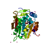
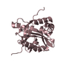
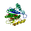
 SIGNALING PROTEIN / alpha-beta-alpha sandwich / phosphopantetheine fold
 Function and homology information

 Authors
 Citation
 Structure visualization
 Downloads & links
 Other downloads


 PDBj
PDBj

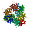
 Assembly


 Mass: 96.063 Da / Num. of mol.: 4 / Source method: obtained synthetically / Formula: SO4
 Mass: 65.409 Da / Num. of mol.: 1 / Source method: obtained synthetically / Formula: Zn
 Mass: 18.015 Da / Num. of mol.: 58 / Source method: isolated from a natural source / Formula: H2O
 Sample preparation
 / Beamline: 19-ID / Wavelength: 1.033 Å
 Processing