 Movie
Movie Controller

[English] 日本語
 Yorodumi
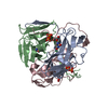
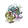
- PDB-1rnj: Crystal structure of inactive mutant dUTPase complexed with subst... -
+ Open data

- Basic information
| Entry | Database: PDB / ID: 1rnj | ||||||
|---|---|---|---|---|---|---|---|
| Title | Crystal structure of inactive mutant dUTPase complexed with substrate analogue imido-dUTP | ||||||
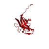
 Components Components | Deoxyuridine 5'-triphosphate nucleotidohydrolase | ||||||
 Keywords Keywords |  HYDROLASE / Jelly Roll / Enzyme-ligand complex HYDROLASE / Jelly Roll / Enzyme-ligand complex | ||||||
| Function / homology |  Function and homology information Function and homology informationdUTP catabolic process / dUMP biosynthetic process / dUTP diphosphatase / dUTP diphosphatase activity / protein homotrimerization / magnesium ion binding / protein-containing complex / identical protein binding / cytosolSimilarity search - Function | ||||||
| Biological species |  Escherichia coli (E. coli) Escherichia coli (E. coli) | ||||||
| Method | X-RAY DIFFRACTION / SYNCHROTRON / MOLECULAR REPLACEMENT / Resolution: 1.7 Å | ||||||
 Authors Authors | Barabas, O. / Pongracz, V. / Kovari, J. / Wilmanns, M. / Vertessy, B.G. | ||||||
 Citation Citation | Journal: J.Biol.Chem. / Year: 2004 Title: Structural Insights into the Catalytic Mechanism of Phosphate Ester Hydrolysis by dUTPase. Authors: Barabas, O. / Pongracz, V. / Kovari, J. / Wilmanns, M. / Vertessy, B.G. #1: Journal: Acta Crystallogr.,Sect.D / Year: 2001Title: Atomic resolution structure of Escherichia coli dUTPase determined ab initio Authors: Gonzalez, A. / Larsson, G. / Persson, R. / Cedergren-Zeppezauer, E. | ||||||
| History |
| ||||||
| Remark 600 | HETEROGEN THE TRIS MOLECULES (TRS), WHICH HAVE INHERENT THREEFOLD SYMMETRY, ARE LOCATED ON A ...HETEROGEN THE TRIS MOLECULES (TRS), WHICH HAVE INHERENT THREEFOLD SYMMETRY, ARE LOCATED ON A CRYSTALLOGRAPHIC THREEFOLD AXIS IN THE PRESENT STRUCTURE. IN AN ASYMMETRIC UNIT THERE IS ONLY ONE THIRD OF THE MOLECULES AND THE COMPLETE MOLECULES ARE BUILT BY THREEFOLD CRYSTALLOGRAPHIC SYMMETRY IN THE CRYSTAL. |
- Structure visualization
| Structure viewer | Molecule: MolmilJmol/JSmol |
|---|
- Downloads & links
-Download
| PDBx/mmCIF format | 1rnj.cif.gz | 50.8 KB | Display | PDBx/mmCIF format |
|---|---|---|---|---|
| PDB format | pdb1rnj.ent.gz | 34.2 KB | Display | PDB format |
| PDBx/mmJSON format | 1rnj.json.gz | Tree view | PDBx/mmJSON format | |
| Others |  Other downloads Other downloads |
-Validation report
| Arichive directory | https://data.pdbj.org/pub/pdb/validation_reports/rn/1rnjftp://data.pdbj.org/pub/pdb/validation_reports/rn/1rnj | HTTPS FTP |
|---|
-Related structure data
| Related structure data | 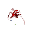 1rn8C 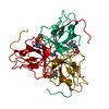 1sehC  1sylC 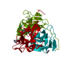 1euwS  1ro1 C: citing same article ( S: Starting model for refinement |
|---|---|
| Similar structure data |


-Links
 PDBj
PDBj
- Assembly
| Deposited unit | 
| |||||||||||||||
|---|---|---|---|---|---|---|---|---|---|---|---|---|---|---|---|---|
| 1 | 
| |||||||||||||||
| Unit cell |
| |||||||||||||||
| Components on special symmetry positions |
| |||||||||||||||
| Details | The biological assembly is a trimer generated from the monomer in the asymmetric unit by the operations: 1-y, x-y, z and 1-x+y, 1-x, z. |
-Components
| #1: Protein | Mass: 16301.631 Da / Num. of mol.: 1 / Mutation: D90N Source method: isolated from a genetically manipulated source Source: (gene. exp.) Escherichia coli (E. coli) / Gene: DUT, DNAS, SOF, B3640 / Plasmid: pET22b / Species (production host): Escherichia coli / Production host: Escherichia coli BL21(DE3) (bacteria) / Strain (production host): BL21(DE3) / References: UniProt: P06968, dUTP diphosphatase | ||
|---|---|---|---|
| #2: Chemical | ChemComp-MG /   Mass: 24.305 Da / Num. of mol.: 1 / Source method: obtained synthetically / Formula: Mg Mass: 24.305 Da / Num. of mol.: 1 / Source method: obtained synthetically / Formula: Mg | ||
| #3: Chemical | ChemComp-DUP / 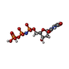  Mass: 467.157 Da / Num. of mol.: 1 / Source method: obtained synthetically / Formula: C9H16N3O13P3 Mass: 467.157 Da / Num. of mol.: 1 / Source method: obtained synthetically / Formula: C9H16N3O13P3 | ||
| #4: Chemical | Tris  Mass: 122.143 Da / Num. of mol.: 2 / Source method: obtained synthetically / Formula: C4H12NO3 / Comment: pH buffer*YM Mass: 122.143 Da / Num. of mol.: 2 / Source method: obtained synthetically / Formula: C4H12NO3 / Comment: pH buffer*YM#5: Water | ChemComp-HOH / | Water Mass: 18.015 Da / Num. of mol.: 221 / Source method: isolated from a natural source / Formula: H2O Mass: 18.015 Da / Num. of mol.: 221 / Source method: isolated from a natural source / Formula: H2O |
-Experimental details
-Experiment
| Experiment | Method: X-RAY DIFFRACTION / Number of used crystals: 1 |
|---|
- Sample preparation
| Crystal | Density Matthews: 2.55 Å3/Da / Density % sol: 51.3 % |
|---|---|
| Crystal grow | Temperature: 293 K / Method: vapor diffusion, hanging drop / pH: 7.8 Details: PEG 3350, sodium acetate, Tris, pH 7.8, VAPOR DIFFUSION, HANGING DROP, temperature 293K |
-Data collection
| Diffraction | Mean temperature: 100 K |
|---|---|
| Diffraction source | Source: SYNCHROTRON / Site: ESRF  / Beamline: ID14-4 / Wavelength: 0.9392 Å / Beamline: ID14-4 / Wavelength: 0.9392 Å |
| Detector | Type: ADSC QUANTUM 4 / Detector: CCD / Date: Mar 8, 2003 |
| Radiation | Monochromator: Si111 crystal / Protocol: SINGLE WAVELENGTH / Monochromatic (M) / Laue (L): M / Scattering type: x-ray |
| Radiation wavelength | Wavelength: 0.9392 Å / Relative weight: 1 |
| Reflection | Resolution: 1.7→22 Å / Num. obs: 18342 / % possible obs: 98.2 % / Observed criterion σ(F): 0 / Observed criterion σ(I): 0 / Redundancy: 20.32 % / Rsym value: 0.059 / Net I/σ(I): 8.4 |
| Reflection shell | Resolution: 1.7→1.74 Å / Redundancy: 20.81 % / Mean I/σ(I) obs: 2 / Num. unique all: 1268 / Rsym value: 0.38 / % possible all: 98.2 |
- Processing
| Software |
| |||||||||||||||||||||||||||||||||||||||||||||||||||||||||||||||||||||||||||||||||||||||||||||||||||||||||
|---|---|---|---|---|---|---|---|---|---|---|---|---|---|---|---|---|---|---|---|---|---|---|---|---|---|---|---|---|---|---|---|---|---|---|---|---|---|---|---|---|---|---|---|---|---|---|---|---|---|---|---|---|---|---|---|---|---|---|---|---|---|---|---|---|---|---|---|---|---|---|---|---|---|---|---|---|---|---|---|---|---|---|---|---|---|---|---|---|---|---|---|---|---|---|---|---|---|---|---|---|---|---|---|---|---|---|
| Refinement | Method to determine structure: MOLECULAR REPLACEMENT Starting model: PDB ENTRY 1EUW Resolution: 1.7→20 Å / Cor.coef. Fo:Fc: 0.967 / Cor.coef. Fo:Fc free: 0.948 / SU B: 1.624 / SU ML: 0.054 / Isotropic thermal model: Isotropic / Cross valid method: THROUGHOUT / σ(F): 0 / ESU R: 0.092 / ESU R Free: 0.091 / Stereochemistry target values: MAXIMUM LIKELIHOOD / Details: HYDROGENS HAVE BEEN ADDED IN THE RIDING POSITIONS
| |||||||||||||||||||||||||||||||||||||||||||||||||||||||||||||||||||||||||||||||||||||||||||||||||||||||||
| Solvent computation | Ion probe radii: 0.8 Å / Shrinkage radii: 0.8 Å / VDW probe radii: 1.4 Å / Solvent model: BABINET MODEL WITH MASK | |||||||||||||||||||||||||||||||||||||||||||||||||||||||||||||||||||||||||||||||||||||||||||||||||||||||||
| Displacement parameters | Biso mean: 19.221 Å2
| |||||||||||||||||||||||||||||||||||||||||||||||||||||||||||||||||||||||||||||||||||||||||||||||||||||||||
| Refinement step | Cycle: LAST / Resolution: 1.7→20 Å
| |||||||||||||||||||||||||||||||||||||||||||||||||||||||||||||||||||||||||||||||||||||||||||||||||||||||||
| Refine LS restraints |
| |||||||||||||||||||||||||||||||||||||||||||||||||||||||||||||||||||||||||||||||||||||||||||||||||||||||||
| LS refinement shell | Resolution: 1.7→1.744 Å / Total num. of bins used: 20
|
