 Movie
Movie Controller

[English] 日本語
 Yorodumi
- PDB-1rla: THREE-DIMENSIONAL STRUCTURE OF RAT LIVER ARGINASE, THE BINUCLEAR ... -
+ Open data

- Basic information
| Entry | Database: PDB / ID: 1rla | ||||||
|---|---|---|---|---|---|---|---|
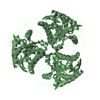
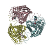
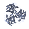
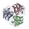
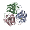
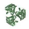
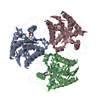
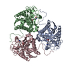
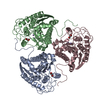
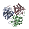
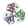
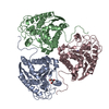
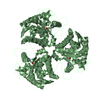
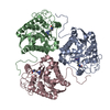
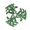
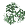
| Title | THREE-DIMENSIONAL STRUCTURE OF RAT LIVER ARGINASE, THE BINUCLEAR MANGANESE METALLOENZYME OF THE UREA CYCLE | ||||||
 Components Components | ARGINASE | ||||||
 Keywords Keywords | HYDROLASE / UREA CYCLE / ARGININE METABOLISM / MAGNESIUM | ||||||
| Function / homology |  Function and homology information Function and homology informationregulation of L-arginine import across plasma membrane / Urea cycle / collagen biosynthetic process / mammary gland involution / positive regulation of neutrophil mediated killing of fungus / arginine metabolic process / negative regulation of T-helper 2 cell cytokine production / response to selenium ion / arginase / arginine catabolic process to ornithine ...regulation of L-arginine import across plasma membrane / Urea cycle / collagen biosynthetic process / mammary gland involution / positive regulation of neutrophil mediated killing of fungus / arginine metabolic process / negative regulation of T-helper 2 cell cytokine production / response to selenium ion / arginase / arginine catabolic process to ornithine / arginase activity / urea cycle / response to methylmercury / response to vitamin A / response to herbicide / response to steroid hormone / response to manganese ion / Neutrophil degranulation / response to zinc ion / negative regulation of type II interferon-mediated signaling pathway / cellular response to glucagon stimulus / defense response to protozoan / response to amine / negative regulation of activated T cell proliferation / response to axon injury / maternal process involved in female pregnancy / response to vitamin E / cellular response to interleukin-4 / response to amino acid / response to cadmium ion / negative regulation of T cell proliferation / cellular response to transforming growth factor beta stimulus / positive regulation of endothelial cell proliferation / cellular response to dexamethasone stimulus / liver development / female pregnancy / lung development / response to peptide hormone / cellular response to hydrogen peroxide / manganese ion binding / adaptive immune response / cellular response to lipopolysaccharide / mitochondrial outer membrane / response to lipopolysaccharide / response to xenobiotic stimulus / innate immune response / neuronal cell body / extracellular space / identical protein binding / cytosol / cytoplasmSimilarity search - Function | ||||||
| Biological species |  Rattus norvegicus (Norway rat) Rattus norvegicus (Norway rat) | ||||||
| Method | X-RAY DIFFRACTION / Resolution: 2.1 Å | ||||||
 Authors Authors | Kanyo, Z. / Scolnick, L. / Ash, D. / Christianson, D.W. | ||||||
 Citation Citation | Journal: Nature / Year: 1996 Title: Structure of a unique binuclear manganese cluster in arginase. Authors: Kanyo, Z.F. / Scolnick, L.R. / Ash, D.E. / Christianson, D.W. #1: Journal: J.Mol.Biol. / Year: 1992Title: Crystallization and Oligomeric Structure of Rat Liver Arginase Authors: Kanyo, Z.F. / Chen, C.Y. / Daghigh, F. / Ash, D.E. / Christianson, D.W. | ||||||
| History |
|
- Structure visualization
| Structure viewer | Molecule: MolmilJmol/JSmol |
|---|
- Downloads & links
-Download
| PDBx/mmCIF format | 1rla.cif.gz | 220.4 KB | Display | PDBx/mmCIF format |
|---|---|---|---|---|
| PDB format | pdb1rla.ent.gz | 178.3 KB | Display | PDB format |
| PDBx/mmJSON format | 1rla.json.gz | Tree view | PDBx/mmJSON format | |
| Others |  Other downloads Other downloads |
-Validation report
| Arichive directory | https://data.pdbj.org/pub/pdb/validation_reports/rl/1rlaftp://data.pdbj.org/pub/pdb/validation_reports/rl/1rla | HTTPS FTP |
|---|
-Related structure data
| Similar structure data |
|---|

-Links
 PDBj
PDBj


- Assembly
| Deposited unit | 
| ||||||||
|---|---|---|---|---|---|---|---|---|---|
| 1 |
| ||||||||
| Unit cell |
|
-Components
| #1: Protein | Mass: 35019.098 Da / Num. of mol.: 3 / Source method: isolated from a natural source / Details: PH 8.5 / Source: (natural) Rattus norvegicus (Norway rat) / Organ: LIVER / References: UniProt: P07824, arginase#2: Chemical | ChemComp-MN /   Mass: 54.938 Da / Num. of mol.: 6 / Source method: obtained synthetically / Formula: Mn Mass: 54.938 Da / Num. of mol.: 6 / Source method: obtained synthetically / Formula: Mn#3: Water | ChemComp-HOH / | Water Mass: 18.015 Da / Num. of mol.: 232 / Source method: isolated from a natural source / Formula: H2O Mass: 18.015 Da / Num. of mol.: 232 / Source method: isolated from a natural source / Formula: H2O |
|---|
-Experimental details
-Experiment
| Experiment | Method: X-RAY DIFFRACTION |
|---|
- Sample preparation
| Crystal | Density Matthews: 2.28 Å3/Da / Density % sol: 48 % | ||||||||||||||||||||||||||||||||||||||||||||||||
|---|---|---|---|---|---|---|---|---|---|---|---|---|---|---|---|---|---|---|---|---|---|---|---|---|---|---|---|---|---|---|---|---|---|---|---|---|---|---|---|---|---|---|---|---|---|---|---|---|---|
| Crystal grow | *PLUS Temperature: 4 ℃ / pH: 8.5 / Method: vapor diffusion, hanging drop / Details: Kanyo, Z.F., (1992) J.Mol.Biol., 224, 1175. | ||||||||||||||||||||||||||||||||||||||||||||||||
| Components of the solutions | *PLUS
|
-Data collection
| Diffraction source | Wavelength: 1.5418 |
|---|---|
| Detector | Type: RIGAKU / Detector: IMAGE PLATE / Date: Sep 17, 1994 |
| Radiation | Monochromatic (M) / Laue (L): M / Scattering type: x-ray |
| Radiation wavelength | Wavelength: 1.5418 Å / Relative weight: 1 |
| Reflection | Rmerge(I) obs: 0.056 |
| Reflection | *PLUS Highest resolution: 2.1 Å / Num. obs: 46162 / % possible obs: 91.6 % / Num. measured all: 147454 |
| Reflection shell | *PLUS % possible obs: 58.3 % / Rmerge(I) obs: 0.365 |
- Processing
| Software |
| |||||||||||||||
|---|---|---|---|---|---|---|---|---|---|---|---|---|---|---|---|---|
| Refinement | Resolution: 2.1→20 Å / Rfactor Rfree: 0.229 / Rfactor Rwork: 0.178 / Rfactor obs: 0.178 / σ(F): 2 | |||||||||||||||
| Refinement step | Cycle: LAST / Resolution: 2.1→20 Å
| |||||||||||||||
| Software | *PLUS Name: X-PLOR / Classification: refinement | |||||||||||||||
| Refinement | *PLUS Num. reflection obs: 47913 | |||||||||||||||
| Solvent computation | *PLUS | |||||||||||||||
| Displacement parameters | *PLUS | |||||||||||||||
| Refine LS restraints | *PLUS
|
