 Movie
Movie Controller

[English] 日本語
 Yorodumi
- PDB-1rd8: Crystal Structure of the 1918 Human H1 Hemagglutinin Precursor (HA0) -
+ Open data

- Basic information
| Entry | Database: PDB / ID: 1rd8 | |||||||||
|---|---|---|---|---|---|---|---|---|---|---|
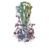
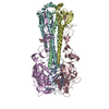
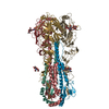
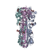
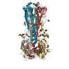
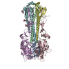
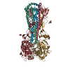
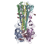
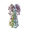
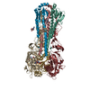
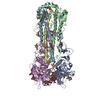
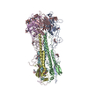
| Title | Crystal Structure of the 1918 Human H1 Hemagglutinin Precursor (HA0) | |||||||||
 Components Components | (hemagglutinin ) x 2 ) x 2 | |||||||||
 Keywords Keywords | VIRAL PROTEIN / GLYCOPROTEIN / MEMBRANE-FUSION PRECURSOR / VIRUS/VIRAL PROTEIN | |||||||||
| Function / homology |  Function and homology informationviral budding from plasma membrane / clathrin-dependent endocytosis of virus by host cell / host cell surface receptor binding / fusion of virus membrane with host plasma membrane / fusion of virus membrane with host endosome membrane / viral envelope / virion attachment to host cell / host cell plasma membrane / virion membrane / membrane Function and homology informationviral budding from plasma membrane / clathrin-dependent endocytosis of virus by host cell / host cell surface receptor binding / fusion of virus membrane with host plasma membrane / fusion of virus membrane with host endosome membrane / viral envelope / virion attachment to host cell / host cell plasma membrane / virion membrane / membraneSimilarity search - Function | |||||||||
| Biological species |   Influenza A virus Influenza A virus | |||||||||
| Method | X-RAY DIFFRACTION / SYNCHROTRON / MOLECULAR REPLACEMENT / Resolution: 3 Å | |||||||||
 Authors Authors | Stevens, J. / Corper, A.L. / Basler, C.F. / Taubenberger, J.K. / Palese, P. / Wilson, I.A. | |||||||||
 Citation Citation | Journal: Science / Year: 2004 Title: Structure of the Uncleaved Human H1 Hemagglutinin from the Extinct 1918 Influenza Virus. Authors: Stevens, J. / Corper, A.L. / Basler, C.F. / Taubenberger, J.K. / Palese, P. / Wilson, I.A. | |||||||||
| History |
|
- Structure visualization
| Structure viewer | Molecule: MolmilJmol/JSmol |
|---|
- Downloads & links
-Download
| PDBx/mmCIF format | 1rd8.cif.gz | 301.4 KB | Display | PDBx/mmCIF format |
|---|---|---|---|---|
| PDB format | pdb1rd8.ent.gz | 249.5 KB | Display | PDB format |
| PDBx/mmJSON format | 1rd8.json.gz | Tree view | PDBx/mmJSON format | |
| Others |  Other downloads Other downloads |
-Validation report
| Arichive directory | https://data.pdbj.org/pub/pdb/validation_reports/rd/1rd8ftp://data.pdbj.org/pub/pdb/validation_reports/rd/1rd8 | HTTPS FTP |
|---|
-Related structure data
| Similar structure data |
|---|
-Links
 PDBj
PDBj


- Assembly
| Deposited unit | 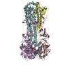
| ||||||||||||||||||||||||||||||||||||||||||||||||||||||||||||||||||||||
|---|---|---|---|---|---|---|---|---|---|---|---|---|---|---|---|---|---|---|---|---|---|---|---|---|---|---|---|---|---|---|---|---|---|---|---|---|---|---|---|---|---|---|---|---|---|---|---|---|---|---|---|---|---|---|---|---|---|---|---|---|---|---|---|---|---|---|---|---|---|---|---|
| 1 |
| ||||||||||||||||||||||||||||||||||||||||||||||||||||||||||||||||||||||
| Unit cell |
| ||||||||||||||||||||||||||||||||||||||||||||||||||||||||||||||||||||||
| Noncrystallographic symmetry (NCS) | NCS domain:
NCS domain segments: Component-ID: 1 / Ens-ID: 1 / Refine code: 0
|
-Components
-Protein , 2 types, 6 molecules ACEBDF
| #1: Protein | Mass: 36971.398 Da / Num. of mol.: 3 / Fragment: Receptor binding domain, HA1 (residues 11-329) Source method: isolated from a genetically manipulated source Source: (gene. exp.) Influenza A virus / Genus: Influenzavirus A / Gene: Hemagglutinin / Plasmid: pAcGP67A / Cell line (production host): SF9 / Production host:   Spodoptera frugiperda (fall armyworm) / References: GenBank: 4325039, UniProt: Q9WFX3*PLUS Spodoptera frugiperda (fall armyworm) / References: GenBank: 4325039, UniProt: Q9WFX3*PLUS#2: Protein | Mass: 20803.992 Da / Num. of mol.: 3 / Fragment: Membrane fusion domain, HA2 (residues 1-175) Source method: isolated from a genetically manipulated source Source: (gene. exp.) Influenza A virus / Genus: Influenzavirus A / Gene: Hemagglutinin / Plasmid: pAcGP67A / Cell line (production host): SF9 / Production host: Spodoptera frugiperda (fall armyworm) / References: GenBank: 4325039, UniProt: Q9WFX3*PLUS |
|---|
-Sugars , 3 types, 9 molecules 
| #3: Polysaccharide | / Mass: 586.542 Da / Num. of mol.: 2 Source method: isolated from a genetically manipulated source #4: Polysaccharide | alpha-D-mannopyranose-(1-3)-beta-D-mannopyranose-(1-4)-2-acetamido-2-deoxy-beta-D-glucopyranose-(1- ...alpha-D-mannopyranose-(1-3)-beta-D-mannopyranose-(1-4)-2-acetamido-2-deoxy-beta-D-glucopyranose-(1-4)-2-acetamido-2-deoxy-beta-D-glucopyranose | / Mass: 748.682 Da / Num. of mol.: 1Source method: isolated from a genetically manipulated source #5: Sugar | ChemComp-NAG / N-Acetylglucosamine Type: D-saccharide, beta linking / Mass: 221.208 Da / Num. of mol.: 6 Type: D-saccharide, beta linking / Mass: 221.208 Da / Num. of mol.: 6Source method: isolated from a genetically manipulated source Formula: C8H15NO6 |
|---|
-Non-polymers , 1 types, 1 molecules 
| #6: Chemical | ChemComp-PO4 / Phosphate Mass: 94.971 Da / Num. of mol.: 1 / Source method: obtained synthetically / Formula: PO4 Mass: 94.971 Da / Num. of mol.: 1 / Source method: obtained synthetically / Formula: PO4 |
|---|
-Experimental details
-Experiment
| Experiment | Method: X-RAY DIFFRACTION / Number of used crystals: 1 |
|---|
- Sample preparation
| Crystal | Density Matthews: 3.9 Å3/Da / Density % sol: 68.46 % | ||||||||||||||||||||||||||||||
|---|---|---|---|---|---|---|---|---|---|---|---|---|---|---|---|---|---|---|---|---|---|---|---|---|---|---|---|---|---|---|---|
| Crystal grow | Temperature: 294 K / Method: vapor diffusion, sitting drop / pH: 5.5 Details: sodium phosphate, potassium phosphate, sodium citrate, pH 5.5, VAPOR DIFFUSION, SITTING DROP, temperature 294K | ||||||||||||||||||||||||||||||
| Crystal grow | *PLUS Method: vapor diffusion, sitting drop | ||||||||||||||||||||||||||||||
| Components of the solutions | *PLUS
|
-Data collection
| Diffraction | Mean temperature: 100 K |
|---|---|
| Diffraction source | Source: SYNCHROTRON / Site: SSRL  / Beamline: BL9-2 / Wavelength: 0.984 Å / Beamline: BL9-2 / Wavelength: 0.984 Å |
| Detector | Type: ADSC QUANTUM 315 / Detector: CCD / Date: Feb 21, 2003 |
| Radiation | Protocol: SINGLE WAVELENGTH / Monochromatic (M) / Laue (L): M / Scattering type: x-ray |
| Radiation wavelength | Wavelength: 0.984 Å / Relative weight: 1 |
| Reflection | Resolution: 3→50 Å / Num. obs: 51346 / % possible obs: 96.4 % / Observed criterion σ(I): 1 / Redundancy: 2.5 % / Rsym value: 0.146 / Net I/σ(I): 7.6 |
| Reflection shell | Resolution: 3→3.07 Å / Redundancy: 2.2 % / Mean I/σ(I) obs: 1.4 / Num. unique all: 3368 / Rsym value: 0.52 / % possible all: 86 |
| Reflection | *PLUS Lowest resolution: 49.4 Å / Num. measured all: 125868 / Rmerge(I) obs: 0.146 |
| Reflection shell | *PLUS Highest resolution: 3 Å / % possible obs: 95.1 % / Num. unique obs: 3368 / Rmerge(I) obs: 0.52 |
- Processing
| Software |
| ||||||||||||||||||||||||||||||||||||||||||||||||||||||||||||||||||||||||||||||||||||||||||||||||||||||||||||||||||||||||||||||||||||||||||||||||||||||||||||||||
|---|---|---|---|---|---|---|---|---|---|---|---|---|---|---|---|---|---|---|---|---|---|---|---|---|---|---|---|---|---|---|---|---|---|---|---|---|---|---|---|---|---|---|---|---|---|---|---|---|---|---|---|---|---|---|---|---|---|---|---|---|---|---|---|---|---|---|---|---|---|---|---|---|---|---|---|---|---|---|---|---|---|---|---|---|---|---|---|---|---|---|---|---|---|---|---|---|---|---|---|---|---|---|---|---|---|---|---|---|---|---|---|---|---|---|---|---|---|---|---|---|---|---|---|---|---|---|---|---|---|---|---|---|---|---|---|---|---|---|---|---|---|---|---|---|---|---|---|---|---|---|---|---|---|---|---|---|---|---|---|---|---|
| Refinement | Method to determine structure: MOLECULAR REPLACEMENT / Resolution: 3→49.39 Å / Cor.coef. Fo:Fc: 0.866 / Cor.coef. Fo:Fc free: 0.824 / SU B: 17.83 / SU ML: 0.323 / Cross valid method: THROUGHOUT / ESU R: 2.586 / ESU R Free: 0.445 / Stereochemistry target values: MAXIMUM LIKELIHOOD Details: The chains were renumbered to be consistent with numbering of the biologically active molecule for which there are existing pdb entries for similar proteins. Renumbering changes made were: ...Details: The chains were renumbered to be consistent with numbering of the biologically active molecule for which there are existing pdb entries for similar proteins. Renumbering changes made were: Segment A 1-503 was split to Chains A 10-329 and Chain B 1-175; Segment B 1-503 was split to Chains C 10-329 and Chain D 1-175; Segment C 1-503 was split to Chains E 10-329 and Chain F 1-175.
| ||||||||||||||||||||||||||||||||||||||||||||||||||||||||||||||||||||||||||||||||||||||||||||||||||||||||||||||||||||||||||||||||||||||||||||||||||||||||||||||||
| Solvent computation | Ion probe radii: 0.8 Å / Shrinkage radii: 0.8 Å / VDW probe radii: 1.4 Å / Solvent model: BABINET MODEL WITH MASK | ||||||||||||||||||||||||||||||||||||||||||||||||||||||||||||||||||||||||||||||||||||||||||||||||||||||||||||||||||||||||||||||||||||||||||||||||||||||||||||||||
| Displacement parameters | Biso mean: 40.312 Å2
| ||||||||||||||||||||||||||||||||||||||||||||||||||||||||||||||||||||||||||||||||||||||||||||||||||||||||||||||||||||||||||||||||||||||||||||||||||||||||||||||||
| Refinement step | Cycle: LAST / Resolution: 3→49.39 Å
| ||||||||||||||||||||||||||||||||||||||||||||||||||||||||||||||||||||||||||||||||||||||||||||||||||||||||||||||||||||||||||||||||||||||||||||||||||||||||||||||||
| Refine LS restraints |
| ||||||||||||||||||||||||||||||||||||||||||||||||||||||||||||||||||||||||||||||||||||||||||||||||||||||||||||||||||||||||||||||||||||||||||||||||||||||||||||||||
| Refine LS restraints NCS | Ens-ID: 1 / Refine-ID: X-RAY DIFFRACTION
| ||||||||||||||||||||||||||||||||||||||||||||||||||||||||||||||||||||||||||||||||||||||||||||||||||||||||||||||||||||||||||||||||||||||||||||||||||||||||||||||||
| LS refinement shell | Resolution: 3→3.079 Å / Total num. of bins used: 20 /
| ||||||||||||||||||||||||||||||||||||||||||||||||||||||||||||||||||||||||||||||||||||||||||||||||||||||||||||||||||||||||||||||||||||||||||||||||||||||||||||||||
| Refinement | *PLUS Highest resolution: 3 Å / Lowest resolution: 49.4 Å / Rfactor Rfree: 0.296 / Rfactor Rwork: 0.27 | ||||||||||||||||||||||||||||||||||||||||||||||||||||||||||||||||||||||||||||||||||||||||||||||||||||||||||||||||||||||||||||||||||||||||||||||||||||||||||||||||
| Solvent computation | *PLUS | ||||||||||||||||||||||||||||||||||||||||||||||||||||||||||||||||||||||||||||||||||||||||||||||||||||||||||||||||||||||||||||||||||||||||||||||||||||||||||||||||
| Displacement parameters | *PLUS | ||||||||||||||||||||||||||||||||||||||||||||||||||||||||||||||||||||||||||||||||||||||||||||||||||||||||||||||||||||||||||||||||||||||||||||||||||||||||||||||||
| Refine LS restraints | *PLUS
|
