 Movie
Movie Controller

[English] 日本語
 Yorodumi
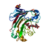
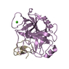
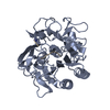
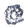
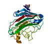
- PDB-1r88: The crystal structure of Mycobacterium tuberculosis MPT51 (FbpC1) -
+ Open data

- Basic information
| Entry | Database: PDB / ID: 1r88 | ||||||
|---|---|---|---|---|---|---|---|
| Title | The crystal structure of Mycobacterium tuberculosis MPT51 (FbpC1) | ||||||
 Components Components | MPT51/MPB51 antigen | ||||||
 Keywords Keywords |  IMMUNE SYSTEM / Mycobacterium tuberculosis / alfa/beta hydrolase fold / antigen 85 / MPT51 / FBPC1 IMMUNE SYSTEM / Mycobacterium tuberculosis / alfa/beta hydrolase fold / antigen 85 / MPT51 / FBPC1 | ||||||
| Function / homology |  Function and homology information Function and homology informationmycolate cell wall layer assembly / zymogen binding / acyltransferase activity, transferring groups other than amino-acyl groups / fibronectin binding / extracellular regionSimilarity search - Function | ||||||
| Biological species |   Mycobacterium tuberculosis (bacteria) Mycobacterium tuberculosis (bacteria) | ||||||
| Method | X-RAY DIFFRACTION / SYNCHROTRON / MOLECULAR REPLACEMENT / Resolution: 1.71 Å | ||||||
 Authors Authors | Wilson, R.A. / Maughan, W.N. / Kremer, L. / Besra, G.S. / Futterer, K. | ||||||
 Citation Citation | Journal: J.Mol.Biol. / Year: 2004 Title: The structure of Mycobacterium tuberculosis MPT51 (FbpC1) defines a new family of non-catalytic alpha/beta hydrolases. Authors: Wilson, R.A. / Maughan, W.N. / Kremer, L. / Besra, G.S. / Futterer, K. | ||||||
| History |
|
- Structure visualization
| Structure viewer | Molecule: MolmilJmol/JSmol |
|---|
- Downloads & links
-Download
| PDBx/mmCIF format | 1r88.cif.gz | 116.6 KB | Display | PDBx/mmCIF format |
|---|---|---|---|---|
| PDB format | pdb1r88.ent.gz | 90.3 KB | Display | PDB format |
| PDBx/mmJSON format | 1r88.json.gz | Tree view | PDBx/mmJSON format | |
| Others |  Other downloads Other downloads |
-Validation report
| Arichive directory | https://data.pdbj.org/pub/pdb/validation_reports/r8/1r88ftp://data.pdbj.org/pub/pdb/validation_reports/r8/1r88 | HTTPS FTP |
|---|
-Related structure data
| Related structure data |  1dqzS S: Starting model for refinement |
|---|---|
| Similar structure data |


-Links
 PDBj
PDBj- Assembly
| Deposited unit | 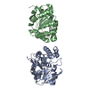
| ||||||||||||||||||
|---|---|---|---|---|---|---|---|---|---|---|---|---|---|---|---|---|---|---|---|
| 1 | 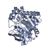
| ||||||||||||||||||
| 2 | 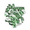
| ||||||||||||||||||
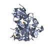
| Unit cell |
| ||||||||||||||||||
| Noncrystallographic symmetry (NCS) | NCS domain:
NCS domain segments: Component-ID: 1 / Ens-ID: 1 / Beg auth comp-ID: ALA / Beg label comp-ID: ALA / End auth comp-ID: GLY / End label comp-ID: GLY / Refine code: 6 / Auth seq-ID: 33 - 281 / Label seq-ID: 8 - 256
| ||||||||||||||||||
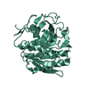
| Details | The biological assembly is a monomer, represented by either of the two molecules in the asymmetric unit |
-Components
| #1: Protein | Mass: 29456.623 Da / Num. of mol.: 2 Source method: isolated from a genetically manipulated source Source: (gene. exp.) Mycobacterium tuberculosis (bacteria) / Gene: fbpC1 / Plasmid: pET23b / Production host: Escherichia coli (E. coli) / Strain (production host): C41(DE3) / References: UniProt: P0A4V6, UniProt: P9WQN7*PLUS#2: Water | ChemComp-HOH / | Water Mass: 18.015 Da / Num. of mol.: 400 / Source method: isolated from a natural source / Formula: H2O Mass: 18.015 Da / Num. of mol.: 400 / Source method: isolated from a natural source / Formula: H2O |
|---|
-Experimental details
-Experiment
| Experiment | Method: X-RAY DIFFRACTION |
|---|
- Sample preparation
| Crystal | Density Matthews: 3.04 Å3/Da / Density % sol: 59.5 % | ||||||||||||||||||||||||||||||||||||||||||
|---|---|---|---|---|---|---|---|---|---|---|---|---|---|---|---|---|---|---|---|---|---|---|---|---|---|---|---|---|---|---|---|---|---|---|---|---|---|---|---|---|---|---|---|
| Crystal grow | *PLUS pH: 4.6 / Method: vapor diffusion, hanging drop | ||||||||||||||||||||||||||||||||||||||||||
| Components of the solutions | *PLUS
|
-Data collection
| Diffraction | Mean temperature: 100 K |
|---|---|
| Diffraction source | Source: SYNCHROTRON / Site: ESRF  / Beamline: ID14-3 / Wavelength: 0.933 Å / Beamline: ID14-3 / Wavelength: 0.933 Å |
| Detector | Type: MARRESEARCH / Detector: CCD / Date: Jun 30, 2003 |
| Radiation | Protocol: SINGLE WAVELENGTH / Monochromatic (M) / Laue (L): M / Scattering type: x-ray |
| Radiation wavelength | Wavelength: 0.933 Å / Relative weight: 1 |
| Reflection | Resolution: 1.7→30 Å / Num. all: 78268 / Num. obs: 78268 / % possible obs: 99.7 % / Observed criterion σ(F): 0 / Observed criterion σ(I): 0 / Redundancy: 6.2 % / Biso Wilson estimate: 18.5 Å2 / Rsym value: 0.077 / Net I/σ(I): 22 |
| Reflection shell | Resolution: 1.7→1.76 Å / Redundancy: 5.7 % / Mean I/σ(I) obs: 3 / Rsym value: 0.489 / % possible all: 99.9 |
| Reflection | *PLUS Highest resolution: 1.7 Å / Num. measured all: 493963 / Rmerge(I) obs: 0.077 |
| Reflection shell | *PLUS % possible obs: 99.9 % / Rmerge(I) obs: 0.489 |
- Processing
| Software |
| ||||||||||||||||||||||||||||||||||||||||||||||||||||||||||||||||||||||||||||||||||||||||||||||||||||
|---|---|---|---|---|---|---|---|---|---|---|---|---|---|---|---|---|---|---|---|---|---|---|---|---|---|---|---|---|---|---|---|---|---|---|---|---|---|---|---|---|---|---|---|---|---|---|---|---|---|---|---|---|---|---|---|---|---|---|---|---|---|---|---|---|---|---|---|---|---|---|---|---|---|---|---|---|---|---|---|---|---|---|---|---|---|---|---|---|---|---|---|---|---|---|---|---|---|---|---|---|---|
| Refinement | Method to determine structure: MOLECULAR REPLACEMENT Starting model: pdb entry 1dqz Resolution: 1.71→30 Å / Cor.coef. Fo:Fc: 0.956 / Cor.coef. Fo:Fc free: 0.949 / SU B: 2.164 / SU ML: 0.068 / Cross valid method: THROUGHOUT / σ(F): 0 / ESU R: 0.097 / ESU R Free: 0.091 / Stereochemistry target values: MAXIMUM LIKELIHOOD / Details: HYDROGENS HAVE BEEN ADDED IN THE RIDING POSITIONS
| ||||||||||||||||||||||||||||||||||||||||||||||||||||||||||||||||||||||||||||||||||||||||||||||||||||
| Solvent computation | Ion probe radii: 0.8 Å / Shrinkage radii: 0.8 Å / VDW probe radii: 1.4 Å / Solvent model: BABINET MODEL WITH MASK | ||||||||||||||||||||||||||||||||||||||||||||||||||||||||||||||||||||||||||||||||||||||||||||||||||||
| Displacement parameters | Biso mean: 12.208 Å2
| ||||||||||||||||||||||||||||||||||||||||||||||||||||||||||||||||||||||||||||||||||||||||||||||||||||
| Refinement step | Cycle: LAST / Resolution: 1.71→30 Å
| ||||||||||||||||||||||||||||||||||||||||||||||||||||||||||||||||||||||||||||||||||||||||||||||||||||
| Refine LS restraints |
| ||||||||||||||||||||||||||||||||||||||||||||||||||||||||||||||||||||||||||||||||||||||||||||||||||||
| Refine LS restraints NCS | Dom-ID: 1 / Auth asym-ID: A / Ens-ID: 1 / Number: 3430 / Refine-ID: X-RAY DIFFRACTION
| ||||||||||||||||||||||||||||||||||||||||||||||||||||||||||||||||||||||||||||||||||||||||||||||||||||
| LS refinement shell | Resolution: 1.706→1.798 Å / Total num. of bins used: 10 /
| ||||||||||||||||||||||||||||||||||||||||||||||||||||||||||||||||||||||||||||||||||||||||||||||||||||
| Refinement TLS params. | Method: refined / Refine-ID: X-RAY DIFFRACTION
| ||||||||||||||||||||||||||||||||||||||||||||||||||||||||||||||||||||||||||||||||||||||||||||||||||||
| Refinement TLS group |
| ||||||||||||||||||||||||||||||||||||||||||||||||||||||||||||||||||||||||||||||||||||||||||||||||||||
| Refinement | *PLUS Highest resolution: 1.7 Å / Lowest resolution: 30 Å / % reflection Rfree: 5 % / Rfactor Rfree: 0.205 / Rfactor Rwork: 0.189 | ||||||||||||||||||||||||||||||||||||||||||||||||||||||||||||||||||||||||||||||||||||||||||||||||||||
| Solvent computation | *PLUS | ||||||||||||||||||||||||||||||||||||||||||||||||||||||||||||||||||||||||||||||||||||||||||||||||||||
| Displacement parameters | *PLUS | ||||||||||||||||||||||||||||||||||||||||||||||||||||||||||||||||||||||||||||||||||||||||||||||||||||
| Refine LS restraints | *PLUS
| ||||||||||||||||||||||||||||||||||||||||||||||||||||||||||||||||||||||||||||||||||||||||||||||||||||
| LS refinement shell | *PLUS Highest resolution: 1.7 Å / Lowest resolution: 1.8 Å |
