[English] 日本語
Yorodumi
- PDB-1r7s: PUTIDAREDOXIN (Fe2S2 ferredoxin), C73G mutant -

+
Open data


ID or keywords:

Loading...

-
Basic information

Entry
Database: PDB / ID: 1r7s
TitlePUTIDAREDOXIN (Fe2S2 ferredoxin), C73G mutant
ComponentsPutidaredoxin
KeywordsELECTRON TRANSFER / CYTOCHROME P450CAM / FERREDOXIN / IRON-SULFUR CLUSTER
Function / homology
Function and homology information


P450-containing electron transport chain / 2 iron, 2 sulfur cluster binding / metal ion binding / cytosol
Similarity search - Function
Adrenodoxin, iron-sulphur binding site / Adrenodoxin family, iron-sulfur binding region signature. / Adrenodoxin / Beta-grasp domain / 2Fe-2S iron-sulfur cluster binding domain / Beta-grasp domain superfamily / 2Fe-2S ferredoxin-type iron-sulfur binding domain profile. / 2Fe-2S ferredoxin-type iron-sulfur binding domain / 2Fe-2S ferredoxin-like superfamily / Ubiquitin-like (UB roll) ...Adrenodoxin, iron-sulphur binding site / Adrenodoxin family, iron-sulfur binding region signature. / Adrenodoxin / Beta-grasp domain / 2Fe-2S iron-sulfur cluster binding domain / Beta-grasp domain superfamily / 2Fe-2S ferredoxin-type iron-sulfur binding domain profile. / 2Fe-2S ferredoxin-type iron-sulfur binding domain / 2Fe-2S ferredoxin-like superfamily / Ubiquitin-like (UB roll) / Roll / Alpha Beta
Similarity search - Domain/homology
FE2/S2 (INORGANIC) CLUSTER / Putidaredoxin
Similarity search - Component
Biological speciesPseudomonas putida (bacteria)
MethodX-RAY DIFFRACTION / SYNCHROTRON / SAD / Resolution: 1.91 Å
AuthorsSmith, N. / Mayhew, M. / Kelly, H. / Robinson, H. / Heroux, A. / Holden, M.J. / Gallagher, D.T.
CitationJournal: Acta Crystallogr.,Sect.D / Year: 2004
Title: Structure of C73G putidaredoxin from Pseudomonas putida.
Authors: Smith, N. / Mayhew, M. / Holden, M.J. / Kelly, H. / Robinson, H. / Heroux, A. / Vilker, V.L. / Gallagher, D.T.
History
DepositionOct 22, 2003Deposition site: RCSB / Processing site: RCSB
Revision 1.0Apr 27, 2004Provider: repository / Type: Initial release
Revision 1.1Apr 29, 2008Group: Version format compliance
Revision 1.2Jul 13, 2011Group: Version format compliance
Revision 1.3Oct 27, 2021Group: Database references / Derived calculations
Category: database_2 / pdbx_struct_conn_angle ...database_2 / pdbx_struct_conn_angle / struct_conn / struct_ref_seq_dif / struct_site
Item: _database_2.pdbx_DOI / _database_2.pdbx_database_accession ..._database_2.pdbx_DOI / _database_2.pdbx_database_accession / _pdbx_struct_conn_angle.ptnr1_auth_asym_id / _pdbx_struct_conn_angle.ptnr1_auth_seq_id / _pdbx_struct_conn_angle.ptnr1_label_asym_id / _pdbx_struct_conn_angle.ptnr1_label_seq_id / _pdbx_struct_conn_angle.ptnr2_auth_asym_id / _pdbx_struct_conn_angle.ptnr2_auth_seq_id / _pdbx_struct_conn_angle.ptnr2_label_asym_id / _pdbx_struct_conn_angle.ptnr3_auth_asym_id / _pdbx_struct_conn_angle.ptnr3_auth_seq_id / _pdbx_struct_conn_angle.ptnr3_label_asym_id / _pdbx_struct_conn_angle.ptnr3_label_seq_id / _pdbx_struct_conn_angle.value / _struct_conn.pdbx_dist_value / _struct_conn.ptnr1_auth_asym_id / _struct_conn.ptnr1_auth_seq_id / _struct_conn.ptnr1_label_asym_id / _struct_conn.ptnr1_label_seq_id / _struct_conn.ptnr2_auth_asym_id / _struct_conn.ptnr2_auth_seq_id / _struct_conn.ptnr2_label_asym_id / _struct_conn.ptnr2_label_atom_id / _struct_ref_seq_dif.details / _struct_site.pdbx_auth_asym_id / _struct_site.pdbx_auth_comp_id / _struct_site.pdbx_auth_seq_id
Revision 1.4Feb 14, 2024Group: Data collection / Category: chem_comp_atom / chem_comp_bond
Remark 300BIOMOLECULE: 1, 2, 3 NUMBER OF BIOMOLECULES IN THIS ENTRY: 3 THIS ENTRY CONTAINS THE ...BIOMOLECULE: 1, 2, 3 NUMBER OF BIOMOLECULES IN THIS ENTRY: 3 THIS ENTRY CONTAINS THE CRYSTALLOGRAPHIC ASYMMETRIC UNIT WHICH CONSISTS OF 3 CHAIN(S). SEE REMARK 350 FOR INFORMATION ON GENERATING THE BIOLOGICAL MOLECULE(S).

-
Structure visualization

Structure viewerMolecule:
MolmilJmol/JSmol

Downloads & links

-
Assembly

Deposited unit
A: Putidaredoxin
B: Putidaredoxin
C: Putidaredoxin
hetero molecules


Theoretical massNumber of molelcules
Total (without water)34,6766
Polymers34,1483
Non-polymers5273
Water5,441302
1
A: Putidaredoxin
hetero molecules


Theoretical massNumber of molelcules
Total (without water)11,5592
Polymers11,3831
Non-polymers1761
Water181
TypeNameSymmetry operationNumber
identity operation1_555x,y,z1
2
B: Putidaredoxin
hetero molecules


Theoretical massNumber of molelcules
Total (without water)11,5592
Polymers11,3831
Non-polymers1761
Water181
TypeNameSymmetry operationNumber
identity operation1_555x,y,z1
3
C: Putidaredoxin
hetero molecules


Theoretical massNumber of molelcules
Total (without water)11,5592
Polymers11,3831
Non-polymers1761
Water181
TypeNameSymmetry operationNumber
identity operation1_555x,y,z1
Unit cell
Length a, b, c (Å)145.076, 78.218, 55.739
Angle α, β, γ (deg.)90.00, 111.33, 90.00
Int Tables number5
Space group name H-MC121
Components on special symmetry positions
IDModelComponents
11A-216-

HOH

Details
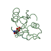
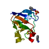
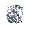
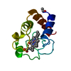
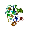
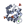
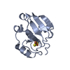
The biological unit is a monomer of 106 residues and one Fe2S2 cluster. This PDB entry (the crystal asymmetric unit) contains 3 independent copies (chains A,B,C).

-
Components

#1: Protein Putidaredoxin / PDX


Mass: 11382.820 Da / Num. of mol.: 3 / Mutation: C73G
Source method: isolated from a genetically manipulated source
Source: (gene. exp.) Pseudomonas putida (bacteria) / Gene: CAMB / Production host: Escherichia coli (E. coli) / References: UniProt: P00259
#2: Chemical ChemComp-FES / FE2/S2 (INORGANIC) CLUSTER / Iron–sulfur cluster


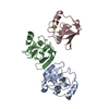
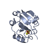
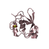
Mass: 175.820 Da / Num. of mol.: 3 / Source method: obtained synthetically / Formula: Fe2S2
#3: Water ChemComp-HOH / water / Water


Mass: 18.015 Da / Num. of mol.: 302 / Source method: isolated from a natural source / Formula: H2O

-
Experimental details

-
Experiment

ExperimentMethod: X-RAY DIFFRACTION / Number of used crystals: 1

-
Sample preparation

CrystalDensity Matthews: 4.31 Å3/Da / Density % sol: 71.47 %
Crystal growTemperature: 277 K / Method: vapor diffusion, hanging drop / pH: 7.5
Details: 1.5M Li2SO4, 0.1M Hepes, 2nM n-octanoyl sucrose, pH 7.5, VAPOR DIFFUSION, HANGING DROP, temperature 277K

-
Data collection

DiffractionMean temperature: 100 K
Diffraction sourceSource: SYNCHROTRON / Site: NSLS / Beamline: X26C / Wavelength: 1.6 Å
DetectorType: ADSC QUANTUM 4 / Detector: CCD / Date: Apr 11, 2003
RadiationMonochromator: SILICON CUT CRYSTAL / Protocol: SINGLE WAVELENGTH / Monochromatic (M) / Laue (L): M / Scattering type: x-ray
Radiation wavelengthWavelength: 1.6 Å / Relative weight: 1
ReflectionResolution: 1.91→30 Å / Num. all: 45116 / Num. obs: 45116 / % possible obs: 100 % / Observed criterion σ(F): 0 / Observed criterion σ(I): 0 / Redundancy: 6.9 % / Biso Wilson estimate: 20.1 Å2 / Rmerge(I) obs: 0.083
Reflection shellResolution: 1.91→1.98 Å / Redundancy: 4.6 % / Rmerge(I) obs: 0.266 / Mean I/σ(I) obs: 6.1 / Num. unique all: 4482 / % possible all: 99.9

-
Processing

Software
NameVersionClassification
CNS1refinement
DENZOdata reduction
SCALEPACKdata scaling
SOLVEphasing
RefinementMethod to determine structure: SAD / Resolution: 1.91→10 Å / Rfactor Rfree error: 0.003 / Data cutoff high absF: 776023.02 / Data cutoff low absF: 0 / Isotropic thermal model: RESTRAINED / Cross valid method: THROUGHOUT / σ(F): 0 / Stereochemistry target values: Engh & Huber
Details: Cluster planarity and coordination geometry were unrestrained.
RfactorNum. reflection% reflectionSelection details
Rfree0.211 2093 5 %RANDOM
Rwork0.188 ---
all0.188 44722 --
obs0.188 42076 97.6 %-
Solvent computationSolvent model: FLAT MODEL / Bsol: 56.2734 Å2 / ksol: 0.514907 e/Å3
Displacement parametersBiso mean: 22.3 Å2
Baniso -1Baniso -2Baniso -3
1--4.41 Å20 Å2-3.59 Å2
2--3.2 Å20 Å2
3---1.21 Å2
Refine analyze
FreeObs
Luzzati coordinate error0.23 Å0.2 Å
Luzzati d res low-5 Å
Luzzati sigma a0.11 Å0.1 Å
Refinement stepCycle: LAST / Resolution: 1.91→10 Å
ProteinNucleic acidLigandSolventTotal
Num. atoms2376 0 12 302 2690
Refine LS restraints
Refine-IDTypeDev ideal
X-RAY DIFFRACTIONc_bond_d0.02
X-RAY DIFFRACTIONc_angle_deg1.9
X-RAY DIFFRACTIONc_dihedral_angle_d26.6
X-RAY DIFFRACTIONc_improper_angle_d1.27
LS refinement shellResolution: 1.91→2.03 Å / Rfactor Rfree error: 0.009 / Total num. of bins used: 6
RfactorNum. reflection% reflection
Rfree0.25 331 5.2 %
Rwork0.23 13085 -
obs-6135 94 %
Xplor file
Refine-IDSerial noParam fileTopol file
X-RAY DIFFRACTION1PROTEIN_REP.PARAMPROTEIN.TOP
X-RAY DIFFRACTION2WATER_REP.PARAMWATER.TOP
X-RAY DIFFRACTION3FES_RELIS.PARAMFES.TOP

+
About Yorodumi

-
News

-
Feb 9, 2022. New format data for meta-information of EMDB entries

New format data for meta-information of EMDB entries

  • Version 3 of the EMDB header file is now the official format.
  • The previous official version 1.9 will be removed from the archive.

Related info.:EMDB header

External links:wwPDB to switch to version 3 of the EMDB data model

-
Aug 12, 2020. Covid-19 info

Covid-19 info

URL: https://pdbj.org/emnavi/covid19.php

New page: Covid-19 featured information page in EM Navigator.

Related info.:Covid-19 info / Mar 5, 2020. Novel coronavirus structure data

+
Mar 5, 2020. Novel coronavirus structure data

Novel coronavirus structure data

Related info.:Yorodumi Speices / Aug 12, 2020. Covid-19 info

External links:COVID-19 featured content - PDBj / Molecule of the Month (242):Coronavirus Proteases

+
Jan 31, 2019. EMDB accession codes are about to change! (news from PDBe EMDB page)

EMDB accession codes are about to change! (news from PDBe EMDB page)

  • The allocation of 4 digits for EMDB accession codes will soon come to an end. Whilst these codes will remain in use, new EMDB accession codes will include an additional digit and will expand incrementally as the available range of codes is exhausted. The current 4-digit format prefixed with “EMD-” (i.e. EMD-XXXX) will advance to a 5-digit format (i.e. EMD-XXXXX), and so on. It is currently estimated that the 4-digit codes will be depleted around Spring 2019, at which point the 5-digit format will come into force.
  • The EM Navigator/Yorodumi systems omit the EMD- prefix.

Related info.:Q: What is EMD? / ID/Accession-code notation in Yorodumi/EM Navigator

External links:EMDB Accession Codes are Changing Soon! / Contact to PDBj

+
Jul 12, 2017. Major update of PDB

Major update of PDB

  • wwPDB released updated PDB data conforming to the new PDBx/mmCIF dictionary.
  • This is a major update changing the version number from 4 to 5, and with Remediation, in which all the entries are updated.
  • In this update, many items about electron microscopy experimental information are reorganized (e.g. em_software).
  • Now, EM Navigator and Yorodumi are based on the updated data.

External links:wwPDB Remediation / Enriched Model Files Conforming to OneDep Data Standards Now Available in the PDB FTP Archive

-
Yorodumi

Thousand views of thousand structures

  • Yorodumi is a browser for structure data from EMDB, PDB, SASBDB, etc.
  • This page is also the successor to EM Navigator detail page, and also detail information page/front-end page for Omokage search.
  • The word "yorodu" (or yorozu) is an old Japanese word meaning "ten thousand". "mi" (miru) is to see.

Related info.:EMDB / PDB / SASBDB / Comparison of 3 databanks / Yorodumi Search / Aug 31, 2016. New EM Navigator & Yorodumi / Yorodumi Papers / Jmol/JSmol / Function and homology information / Changes in new EM Navigator and Yorodumi

Read more