 Movie
Movie Controller

[English] 日本語
 Yorodumi
- PDB-1r5j: Crystal Structure of a Phosphotransacetylase from Streptococcus p... -
+ Open data

- Basic information
| Entry | Database: PDB / ID: 1r5j | ||||||
|---|---|---|---|---|---|---|---|
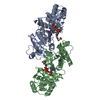
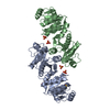
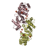
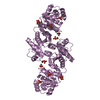
| Title | Crystal Structure of a Phosphotransacetylase from Streptococcus pyogenes | ||||||
 Components Components | putative phosphotransacetylase Phosphate acetyltransferase Phosphate acetyltransferase | ||||||
 Keywords Keywords | TRANSFERASE / lactate dehydrogenase-like nucleotide-binding fold / Structural Genomics / BSGC structure funded by NIH / Protein Structure Initiative / PSI / Berkeley Structural Genomics Center | ||||||
| Function / homology |  Function and homology information Function and homology information | ||||||
| Biological species |  Streptococcus pyogenes (bacteria) Streptococcus pyogenes (bacteria) | ||||||
| Method | X-RAY DIFFRACTION / SYNCHROTRON / MAD / Resolution: 2.7 Å | ||||||
 Authors Authors | Xu, Q.S. / Shin, D.H. / Pufan, R. / Yokota, H. / Kim, R. / Kim, S.H. / Berkeley Structural Genomics Center (BSGC) | ||||||
 Citation Citation | Journal: Proteins / Year: 2004 Title: Crystal structure of a phosphotransacetylase from Streptococcus pyogenes. Authors: Xu, Q.S. / Shin, D.H. / Pufan, R. / Yokota, H. / Kim, R. / Kim, S.H. | ||||||
| History |
|
- Structure visualization
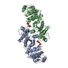
| Structure viewer | Molecule: MolmilJmol/JSmol |
|---|
- Downloads & links
-Download
| PDBx/mmCIF format | 1r5j.cif.gz | 132.6 KB | Display | PDBx/mmCIF format |
|---|---|---|---|---|
| PDB format | pdb1r5j.ent.gz | 111 KB | Display | PDB format |
| PDBx/mmJSON format | 1r5j.json.gz | Tree view | PDBx/mmJSON format | |
| Others |  Other downloads Other downloads |
-Validation report
| Arichive directory | https://data.pdbj.org/pub/pdb/validation_reports/r5/1r5jftp://data.pdbj.org/pub/pdb/validation_reports/r5/1r5j | HTTPS FTP |
|---|
-Related structure data
| Similar structure data | |
|---|---|
| Other databases |


-Links
 PDBj
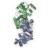
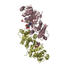
PDBj- Assembly
| Deposited unit | 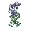
| ||||||||
|---|---|---|---|---|---|---|---|---|---|
| 1 |
| ||||||||
| 2 | 
| ||||||||
| Unit cell |
|
-Components
| #1: Protein | Phosphate acetyltransferase Mass: 36673.215 Da / Num. of mol.: 2 Source method: isolated from a genetically manipulated source Source: (gene. exp.) Streptococcus pyogenes (bacteria) / Plasmid: pLIC.B4 / Production host: Escherichia coli (E. coli) / Strain (production host): BL21(DE3)/pSJS1244 / References: UniProt: Q99ZQ5, phosphate acetyltransferase#2: Water | ChemComp-HOH / | Water Mass: 18.015 Da / Num. of mol.: 30 / Source method: isolated from a natural source / Formula: H2O Mass: 18.015 Da / Num. of mol.: 30 / Source method: isolated from a natural source / Formula: H2O |
|---|
-Experimental details
-Experiment
| Experiment | Method: X-RAY DIFFRACTION / Number of used crystals: 1 |
|---|
- Sample preparation
| Crystal | Density Matthews: 3.05 Å3/Da / Density % sol: 59.6 % |
|---|---|
| Crystal grow | Temperature: 298 K / pH: 5.5 Details: BisTris, PEG 3350, sodium chloride, pH 5.5, VAPOR DIFFUSION, HANGING DROP, temperature 298K, pH 5.50 |
-Data collection
| Diffraction | Mean temperature: 100 K | ||||||||||||
|---|---|---|---|---|---|---|---|---|---|---|---|---|---|
| Diffraction source | Source: SYNCHROTRON / Site: ALS  / Beamline: 5.0.2 / Wavelength: 0.9797, 0.9794, 0.9500 / Beamline: 5.0.2 / Wavelength: 0.9797, 0.9794, 0.9500 | ||||||||||||
| Detector | Type: ADSC QUANTUM 4 / Detector: CCD / Date: Jun 28, 2003 / Details: MIRRORS | ||||||||||||
| Radiation | Monochromator: DOUBLE-CRYSTAL, SI(111) / Protocol: MAD / Monochromatic (M) / Laue (L): M / Scattering type: x-ray | ||||||||||||
| Radiation wavelength |
| ||||||||||||
| Reflection | Resolution: 2.7→20 Å / Num. obs: 25144 / % possible obs: 100 % / Observed criterion σ(I): -3 / Redundancy: 23.2 % / Biso Wilson estimate: 69.9 Å2 / Rsym value: 0.119 / Net I/σ(I): 23.9 | ||||||||||||
| Reflection shell | Resolution: 2.7→2.75 Å / Mean I/σ(I) obs: 1.3 / % possible all: 100 |
- Processing
| Software |
| ||||||||||||||||||||||||||||||||||||||||||||||||||||||||||||||||||||||||||||||||
|---|---|---|---|---|---|---|---|---|---|---|---|---|---|---|---|---|---|---|---|---|---|---|---|---|---|---|---|---|---|---|---|---|---|---|---|---|---|---|---|---|---|---|---|---|---|---|---|---|---|---|---|---|---|---|---|---|---|---|---|---|---|---|---|---|---|---|---|---|---|---|---|---|---|---|---|---|---|---|---|---|---|
| Refinement | Method to determine structure: MAD / Resolution: 2.7→19.93 Å / Rfactor Rfree error: 0.006 / Data cutoff high absF: 3842196.04 / Data cutoff low absF: 0 / Isotropic thermal model: RESTRAINED / Cross valid method: THROUGHOUT / σ(F): 0 / Stereochemistry target values: ENGH & HUBER
| ||||||||||||||||||||||||||||||||||||||||||||||||||||||||||||||||||||||||||||||||
| Solvent computation | Solvent model: FLAT MODEL / Bsol: 38.4 Å2 / ksol: 0.29 e/Å3 | ||||||||||||||||||||||||||||||||||||||||||||||||||||||||||||||||||||||||||||||||
| Displacement parameters | Biso mean: 72.5 Å2
| ||||||||||||||||||||||||||||||||||||||||||||||||||||||||||||||||||||||||||||||||
| Refine analyze |
| ||||||||||||||||||||||||||||||||||||||||||||||||||||||||||||||||||||||||||||||||
| Refinement step | Cycle: LAST / Resolution: 2.7→19.93 Å
| ||||||||||||||||||||||||||||||||||||||||||||||||||||||||||||||||||||||||||||||||
| Refine LS restraints |
| ||||||||||||||||||||||||||||||||||||||||||||||||||||||||||||||||||||||||||||||||
| LS refinement shell | Resolution: 2.7→2.8 Å / Rfactor Rfree error: 0.028 / Total num. of bins used: 10
| ||||||||||||||||||||||||||||||||||||||||||||||||||||||||||||||||||||||||||||||||
| Xplor file |
|
