 Movie
Movie Controller

[English] 日本語
 Yorodumi
- PDB-1r45: ADP-ribosyltransferase C3bot2 from Clostridium botulinum, triclin... -
+ Open data

- Basic information
| Entry | Database: PDB / ID: 1r45 | ||||||
|---|---|---|---|---|---|---|---|
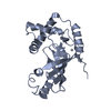
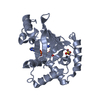
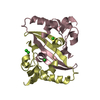
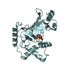
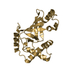
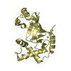
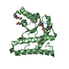
| Title | ADP-ribosyltransferase C3bot2 from Clostridium botulinum, triclinic form | ||||||
 Components Components | Mono-ADP-ribosyltransferase C3 | ||||||
 Keywords Keywords |  TRANSFERASE / ADP-RIBOSYLTRANSFERASE / BINARY TOXIN / C3 EXOENZYME TRANSFERASE / ADP-RIBOSYLTRANSFERASE / BINARY TOXIN / C3 EXOENZYME | ||||||
| Function / homology |  Function and homology information Function and homology informationNAD+-protein ADP-ribosyltransferase activity / Transferases; Glycosyltransferases; Pentosyltransferases / nucleotidyltransferase activity / extracellular regionSimilarity search - Function | ||||||
| Biological species |  Clostridium phage c-st (virus) Clostridium phage c-st (virus) | ||||||
| Method | X-RAY DIFFRACTION / SYNCHROTRON / MOLECULAR REPLACEMENT / Resolution: 1.57 Å | ||||||
 Authors Authors | Teplyakov, A. / Obmolova, G. / Gilliland, G.L. / Narumiya, S. | ||||||
 Citation Citation | Journal: To be Published Title: Crystal structure of ADP-ribosyltransferase C3bot2 from Clostridium botulinum Authors: Teplyakov, A. / Obmolova, G. / Gilliland, G.L. / Narumiya, S. | ||||||
| History |
|
- Structure visualization
| Structure viewer | Molecule: MolmilJmol/JSmol |
|---|
- Downloads & links
-Download
| PDBx/mmCIF format | 1r45.cif.gz | 190 KB | Display | PDBx/mmCIF format |
|---|---|---|---|---|
| PDB format | pdb1r45.ent.gz | 150.5 KB | Display | PDB format |
| PDBx/mmJSON format | 1r45.json.gz | Tree view | PDBx/mmJSON format | |
| Others |  Other downloads Other downloads |
-Validation report
| Arichive directory | https://data.pdbj.org/pub/pdb/validation_reports/r4/1r45ftp://data.pdbj.org/pub/pdb/validation_reports/r4/1r45 | HTTPS FTP |
|---|
-Related structure data
| Related structure data | 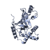 1g24S S: Starting model for refinement |
|---|---|
| Similar structure data |


-Links
 PDBj
PDBj- Assembly
| Deposited unit | 
| ||||||||
|---|---|---|---|---|---|---|---|---|---|
| 1 | 
| ||||||||
| 2 | 
| ||||||||
| 3 | 
| ||||||||
| 4 | 
| ||||||||
| 5 | 
| ||||||||
| Unit cell |
|
-Components
| #1: Protein | Mass: 23225.521 Da / Num. of mol.: 4 / Fragment: mature protein Source method: isolated from a genetically manipulated source Source: (gene. exp.) Clostridium phage c-st (virus) / Strain: c003-9 / Plasmid: pET3a / Species (production host): Escherichia coli / Production host:  Escherichia coli BL21(DE3) (bacteria) / Strain (production host): BL21 (DE3) Escherichia coli BL21(DE3) (bacteria) / Strain (production host): BL21 (DE3)References: UniProt: Q00901, Transferases; Glycosyltransferases; Pentosyltransferases#2: Chemical | ChemComp-SO4 / Sulfate  Mass: 96.063 Da / Num. of mol.: 8 / Source method: obtained synthetically / Formula: SO4 Mass: 96.063 Da / Num. of mol.: 8 / Source method: obtained synthetically / Formula: SO4#3: Chemical | Glycerol  Mass: 92.094 Da / Num. of mol.: 2 / Source method: obtained synthetically / Formula: C3H8O3 Mass: 92.094 Da / Num. of mol.: 2 / Source method: obtained synthetically / Formula: C3H8O3#4: Water | ChemComp-HOH / | Water Mass: 18.015 Da / Num. of mol.: 907 / Source method: isolated from a natural source / Formula: H2O Mass: 18.015 Da / Num. of mol.: 907 / Source method: isolated from a natural source / Formula: H2O |
|---|
-Experimental details
-Experiment
| Experiment | Method: X-RAY DIFFRACTION / Number of used crystals: 1 |
|---|
- Sample preparation
| Crystal | Density Matthews: 2.6 Å3/Da / Density % sol: 52 % |
|---|---|
| Crystal grow | Temperature: 295 K / Method: vapor diffusion, hanging drop / pH: 7.5 Details: 0.2 M Lithium Sulfate, 7% PEG 4000, 0.1 M HEPES, pH 7.5, VAPOR DIFFUSION, HANGING DROP, temperature 295K |
-Data collection
| Diffraction | Mean temperature: 100 K |
|---|---|
| Diffraction source | Source: SYNCHROTRON / Site: EMBL/DESY, HAMBURG  / Beamline: BW7B / Wavelength: 1.1 Å / Beamline: BW7B / Wavelength: 1.1 Å |
| Detector | Type: MARRESEARCH / Detector: IMAGE PLATE / Date: May 9, 1997 / Details: mirror |
| Radiation | Monochromator: Germanium / Protocol: SINGLE WAVELENGTH / Monochromatic (M) / Laue (L): M / Scattering type: x-ray |
| Radiation wavelength | Wavelength: 1.1 Å / Relative weight: 1 |
| Reflection | Resolution: 1.57→20 Å / Num. all: 118341 / Num. obs: 118341 / % possible obs: 94 % / Observed criterion σ(F): 0 / Observed criterion σ(I): -3 / Redundancy: 2.1 % / Biso Wilson estimate: 14 Å2 / Rmerge(I) obs: 0.034 / Net I/σ(I): 25.6 |
| Reflection shell | Resolution: 1.57→1.6 Å / Redundancy: 1.7 % / Rmerge(I) obs: 0.068 / Mean I/σ(I) obs: 12.4 / % possible all: 90 |
- Processing
| Software |
| ||||||||||||||||||||||||||||||||||||||||||||||||||||||||||||||||||||||
|---|---|---|---|---|---|---|---|---|---|---|---|---|---|---|---|---|---|---|---|---|---|---|---|---|---|---|---|---|---|---|---|---|---|---|---|---|---|---|---|---|---|---|---|---|---|---|---|---|---|---|---|---|---|---|---|---|---|---|---|---|---|---|---|---|---|---|---|---|---|---|---|
| Refinement | Method to determine structure: MOLECULAR REPLACEMENT Starting model: 1G24-A Resolution: 1.57→15 Å / Cor.coef. Fo:Fc: 0.956 / Cor.coef. Fo:Fc free: 0.939 / SU B: 1.208 / SU ML: 0.045 / Cross valid method: THROUGHOUT / σ(F): 0 / ESU R: 0.08 / ESU R Free: 0.083
| ||||||||||||||||||||||||||||||||||||||||||||||||||||||||||||||||||||||
| Solvent computation | Ion probe radii: 0.8 Å / Shrinkage radii: 0.8 Å / VDW probe radii: 1.4 Å / Solvent model: BABINET MODEL WITH MASK | ||||||||||||||||||||||||||||||||||||||||||||||||||||||||||||||||||||||
| Displacement parameters | Biso mean: 18.3 Å2
| ||||||||||||||||||||||||||||||||||||||||||||||||||||||||||||||||||||||
| Refine analyze | Luzzati coordinate error obs: 0.08 Å | ||||||||||||||||||||||||||||||||||||||||||||||||||||||||||||||||||||||
| Refinement step | Cycle: LAST / Resolution: 1.57→15 Å
| ||||||||||||||||||||||||||||||||||||||||||||||||||||||||||||||||||||||
| Refine LS restraints |
| ||||||||||||||||||||||||||||||||||||||||||||||||||||||||||||||||||||||
| LS refinement shell | Resolution: 1.57→1.608 Å / Total num. of bins used: 20 /
|
