 Movie
Movie Controller


+ Open data

- Basic information
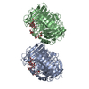
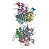
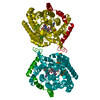
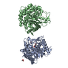
| Entry | Database: PDB / ID: 1qzq | ||||||
|---|---|---|---|---|---|---|---|
| Title | human Tyrosyl DNA phosphodiesterase | ||||||
 Components Components | tyrosyl-DNA phosphodiesterase 1 | ||||||
 Keywords Keywords |  hydrolase / DNA binding protein / DNA Repair / replication hydrolase / DNA binding protein / DNA Repair / replication | ||||||
| Function / homology |  Function and homology information Function and homology information3'-tyrosyl-DNA phosphodiesterase activity / single strand break repair / Hydrolases; Acting on ester bonds; Phosphoric-diester hydrolases / exonuclease activity / Nonhomologous End-Joining (NHEJ) / double-strand break repair / single-stranded DNA binding / double-stranded DNA binding / intracellular membrane-bounded organelle / DNA repair ...3'-tyrosyl-DNA phosphodiesterase activity / single strand break repair / Hydrolases; Acting on ester bonds; Phosphoric-diester hydrolases / exonuclease activity / Nonhomologous End-Joining (NHEJ) / double-strand break repair / single-stranded DNA binding / double-stranded DNA binding / intracellular membrane-bounded organelle / DNA repair / nucleoplasm / nucleus / plasma membrane / cytoplasmSimilarity search - Function | ||||||
| Biological species |  Homo sapiens (human) Homo sapiens (human) | ||||||
| Method | X-RAY DIFFRACTION / SYNCHROTRON / MOLECULAR REPLACEMENT / Resolution: 2.4 Å | ||||||
 Authors Authors | Raymond, A.C. / Rideout, M.C. / Staker, B. / Hjerrild, K. / Burgin Jr., A.B. | ||||||
 Citation Citation | Journal: J.Mol.Biol. / Year: 2004 Title: Analysis of Human Tyrosyl-DNA Phosphodiesterase I Catalytic Residues. Authors: Raymond, A.C. / Rideout, M.C. / Staker, B. / Hjerrild, K. / Burgin Jr., A.B. | ||||||
| History |
|
- Structure visualization
| Structure viewer | Molecule: MolmilJmol/JSmol |
|---|
- Downloads & links
-Download
| PDBx/mmCIF format | 1qzq.cif.gz | 189.4 KB | Display | PDBx/mmCIF format |
|---|---|---|---|---|
| PDB format | pdb1qzq.ent.gz | 150.2 KB | Display | PDB format |
| PDBx/mmJSON format | 1qzq.json.gz | Tree view | PDBx/mmJSON format | |
| Others |  Other downloads Other downloads |
-Validation report
| Arichive directory | https://data.pdbj.org/pub/pdb/validation_reports/qz/1qzqftp://data.pdbj.org/pub/pdb/validation_reports/qz/1qzq | HTTPS FTP |
|---|
-Related structure data
| Similar structure data |
|---|


-Links
 PDBj
PDBj

- Assembly
| Deposited unit | 
| ||||||||
|---|---|---|---|---|---|---|---|---|---|
| 1 |
| ||||||||
| Unit cell |
|
-Components
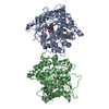
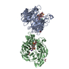
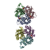
| #1: Protein | Mass: 54919.340 Da / Num. of mol.: 2 / Fragment: residues 149-608 Source method: isolated from a genetically manipulated source Source: (gene. exp.) Homo sapiens (human) / Gene: tdp1 / Production host:  Escherichia coli (E. coli) / References: UniProt: Q9NUW8 Escherichia coli (E. coli) / References: UniProt: Q9NUW8#2: Water | ChemComp-HOH / | Water Mass: 18.015 Da / Num. of mol.: 238 / Source method: isolated from a natural source / Formula: H2O Mass: 18.015 Da / Num. of mol.: 238 / Source method: isolated from a natural source / Formula: H2O |
|---|
-Experimental details
-Experiment
| Experiment | Method: X-RAY DIFFRACTION / Number of used crystals: 1 |
|---|
- Sample preparation
| Crystal | Density Matthews: 2.32 Å3/Da / Density % sol: 47.04 % |
|---|---|
| Crystal grow | Temperature: 289 K / Method: vapor diffusion, sitting drop / pH: 9.4 Details: PEG 8000, CHES, 8mM spermine, pH 9.4, VAPOR DIFFUSION, SITTING DROP, temperature 16K |
-Data collection
| Diffraction source | Source: SYNCHROTRON / Site: APS  / Beamline: 32-ID / Wavelength: 1 Å / Beamline: 32-ID / Wavelength: 1 Å |
|---|---|
| Detector | Type: MARRESEARCH / Detector: CCD |
| Radiation | Protocol: SINGLE WAVELENGTH / Monochromatic (M) / Laue (L): M / Scattering type: x-ray |
| Radiation wavelength | Wavelength: 1 Å / Relative weight: 1 |
| Reflection | Resolution: 2.4→50 Å / Num. obs: 40794 / % possible obs: 99.4 % / Observed criterion σ(I): 0 / Redundancy: 6.1 % / Biso Wilson estimate: 26.7 Å2 / Rsym value: 0.097 / Net I/σ(I): 18.5 |
| Reflection shell | Resolution: 2.4→2.49 Å / Redundancy: 5.8 % / Mean I/σ(I) obs: 3.6 / Num. unique all: 6326 / Rsym value: 0.403 |
- Processing
| Software |
| ||||||||||||||||||||||||||||||||||||||||||||||||||||||||||||
|---|---|---|---|---|---|---|---|---|---|---|---|---|---|---|---|---|---|---|---|---|---|---|---|---|---|---|---|---|---|---|---|---|---|---|---|---|---|---|---|---|---|---|---|---|---|---|---|---|---|---|---|---|---|---|---|---|---|---|---|---|---|
| Refinement | Method to determine structure: MOLECULAR REPLACEMENT / Resolution: 2.4→48.54 Å / Rfactor Rfree error: 0.005 / Data cutoff high absF: 2371494.44 / Data cutoff high rms absF: 2371494.44 / Data cutoff low absF: 0 / Isotropic thermal model: RESTRAINED / Cross valid method: THROUGHOUT / σ(F): 0
| ||||||||||||||||||||||||||||||||||||||||||||||||||||||||||||
| Solvent computation | Solvent model: FLAT MODEL / Bsol: 32.301 Å2 / ksol: 0.367093 e/Å3 | ||||||||||||||||||||||||||||||||||||||||||||||||||||||||||||
| Displacement parameters | Biso mean: 30.9 Å2
| ||||||||||||||||||||||||||||||||||||||||||||||||||||||||||||
| Refine analyze |
| ||||||||||||||||||||||||||||||||||||||||||||||||||||||||||||
| Refinement step | Cycle: LAST / Resolution: 2.4→48.54 Å
| ||||||||||||||||||||||||||||||||||||||||||||||||||||||||||||
| Refine LS restraints |
| ||||||||||||||||||||||||||||||||||||||||||||||||||||||||||||
| LS refinement shell | Resolution: 2.4→2.55 Å / Rfactor Rfree error: 0.016 / Total num. of bins used: 6
| ||||||||||||||||||||||||||||||||||||||||||||||||||||||||||||
| Xplor file |
|
