 Movie
Movie Controller


+ Open data

- Basic information
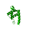
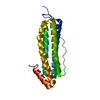
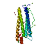
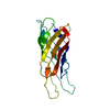
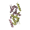
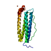
| Entry | Database: PDB / ID: 1qxx | ||||||
|---|---|---|---|---|---|---|---|
| Title | CRYSTAL STRUCTURE OF THE C-TERMINAL DOMAIN OF TONB | ||||||
 Components Components | TonB protein | ||||||
 Keywords Keywords |  TRANSPORT PROTEIN / TonB Dimerization TRANSPORT PROTEIN / TonB Dimerization | ||||||
| Function / homology |  Function and homology information Function and homology informationreceptor-mediated bacteriophage irreversible attachment to host cell / colicin transport / energy transducer activity / cell envelope / cobalamin transport / siderophore transport / intracellular monoatomic cation homeostasis / plasma membrane protein complex / transmembrane transporter complex / cell outer membrane ...receptor-mediated bacteriophage irreversible attachment to host cell / colicin transport / energy transducer activity / cell envelope / cobalamin transport / siderophore transport / intracellular monoatomic cation homeostasis / plasma membrane protein complex / transmembrane transporter complex / cell outer membrane / transmembrane transport / protein transport / outer membrane-bounded periplasmic space / intracellular iron ion homeostasis / protein domain specific binding / membrane / plasma membraneSimilarity search - Function | ||||||
| Biological species |  Escherichia coli (E. coli) Escherichia coli (E. coli) | ||||||
| Method | X-RAY DIFFRACTION / SYNCHROTRON / MOLECULAR REPLACEMENT / Resolution: 2.7 Å | ||||||
 Authors Authors | Koedding, J. / Howard, P. / Kaufmann, L. / Polzer, P. / Lustig, A. / Welte, W. | ||||||
 Citation Citation | Journal: J.Biol.Chem. / Year: 2004 Title: Dimerization of TonB is not essential for its binding to the outer membrane siderophore receptor FhuA of Escherichia coli. Authors: Koedding, J. / Howard, P. / Kaufmann, L. / Polzer, P. / Lustig, A. / Welte, W. | ||||||
| History |
|
- Structure visualization
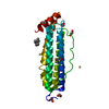
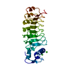
| Structure viewer | Molecule: MolmilJmol/JSmol |
|---|
- Downloads & links
-Download
| PDBx/mmCIF format | 1qxx.cif.gz | 27.1 KB | Display | PDBx/mmCIF format |
|---|---|---|---|---|
| PDB format | pdb1qxx.ent.gz | 17.9 KB | Display | PDB format |
| PDBx/mmJSON format | 1qxx.json.gz | Tree view | PDBx/mmJSON format | |
| Others |  Other downloads Other downloads |
-Validation report
| Arichive directory | https://data.pdbj.org/pub/pdb/validation_reports/qx/1qxxftp://data.pdbj.org/pub/pdb/validation_reports/qx/1qxx | HTTPS FTP |
|---|
-Related structure data
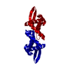
| Related structure data | 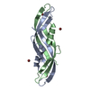 1ihrS S: Starting model for refinement |
|---|---|
| Similar structure data |

-Links
 PDBj
PDBj- Assembly
| Deposited unit | 
| ||||||||
|---|---|---|---|---|---|---|---|---|---|
| 1 | 
| ||||||||
| Unit cell |
| ||||||||
| Details | The second part of the dimer is generated by symmetry operations in P6422 |
-Components
| #1: Protein | Mass: 8563.893 Da / Num. of mol.: 1 / Fragment: residues 164-239 Source method: isolated from a genetically manipulated source Source: (gene. exp.) Escherichia coli (E. coli) / Gene: TONB / Plasmid: pET30a / Species (production host): Escherichia coli / Production host: Escherichia coli BL21(DE3) (bacteria) / Strain (production host): BL21(DE3) / References: UniProt: P02929 |
|---|---|
| #2: Water | ChemComp-HOH / Water Mass: 18.015 Da / Num. of mol.: 31 / Source method: isolated from a natural source / Formula: H2O Mass: 18.015 Da / Num. of mol.: 31 / Source method: isolated from a natural source / Formula: H2O |
-Experimental details
-Experiment
| Experiment | Method: X-RAY DIFFRACTION / Number of used crystals: 1 |
|---|
- Sample preparation
| Crystal | Density Matthews: 3.89 Å3/Da / Density % sol: 68.42 % |
|---|---|
| Crystal grow | Temperature: 301 K / Method: vapor diffusion, hanging drop / pH: 5.6 Details: Sodium formiate, sodium citrate, pH 5.6, VAPOR DIFFUSION, HANGING DROP, temperature 301K |
-Data collection
| Diffraction | Mean temperature: 100 K |
|---|---|
| Diffraction source | Source: SYNCHROTRON / Site: ESRF  / Beamline: ID14-4 / Wavelength: 0.934 Å / Beamline: ID14-4 / Wavelength: 0.934 Å |
| Detector | Type: ADSC QUANTUM 4 / Detector: CCD / Date: Nov 22, 2002 |
| Radiation | Monochromator: Si 111 CHANNEL / Protocol: SINGLE WAVELENGTH / Monochromatic (M) / Laue (L): M / Scattering type: x-ray |
| Radiation wavelength | Wavelength: 0.934 Å / Relative weight: 1 |
| Reflection | Resolution: 2.7→26.44 Å / Num. all: 5019 / Num. obs: 4052 / % possible obs: 97.5 % / Observed criterion σ(F): 0 / Observed criterion σ(I): 0 / Redundancy: 9.68 % / Rmerge(I) obs: 0.049 / Rsym value: 0.049 / Net I/σ(I): 19.32 |
| Reflection shell | Resolution: 2.7→2.8 Å / Redundancy: 10.1 % / Rmerge(I) obs: 0.463 / Mean I/σ(I) obs: 3.56 / Num. unique all: 409 / Rsym value: 0.463 / % possible all: 97.6 |
- Processing
| Software |
| |||||||||||||||||||||||||
|---|---|---|---|---|---|---|---|---|---|---|---|---|---|---|---|---|---|---|---|---|---|---|---|---|---|---|
| Refinement | Method to determine structure: MOLECULAR REPLACEMENT Starting model: 1IHR Resolution: 2.7→26.44 Å / σ(F): 0 / σ(I): 0 / Stereochemistry target values: Engh & Huber
| |||||||||||||||||||||||||
| Refinement step | Cycle: LAST / Resolution: 2.7→26.44 Å
| |||||||||||||||||||||||||
| Refine LS restraints |
|
