 Movie
Movie Controller

[English] 日本語
 Yorodumi
- PDB-1qsm: Histone Acetyltransferase HPA2 from Saccharomyces Cerevisiae in C... -
+ Open data

- Basic information
| Entry | Database: PDB / ID: 1qsm | ||||||
|---|---|---|---|---|---|---|---|
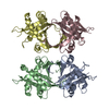
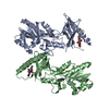
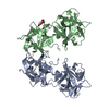
| Title | Histone Acetyltransferase HPA2 from Saccharomyces Cerevisiae in Complex with Acetyl Coenzyme A | ||||||
 Components Components | HPA2 HISTONE ACETYLTRANSFERASE | ||||||
 Keywords Keywords |  TRANSFERASE / PROTEIN-ACETYL COENZYME A COMPLEX / ACETYLTRANSFERASE TRANSFERASE / PROTEIN-ACETYL COENZYME A COMPLEX / ACETYLTRANSFERASE | ||||||
| Function / homology |  Function and homology information Function and homology informationpolyamine acetylation / Hpa2 acetyltransferase complex / N-acetyltransferase activity / protein acetylation / histone acetyltransferase activity / histone acetyltransferase / identical protein binding / nucleus / cytoplasmSimilarity search - Function | ||||||
| Biological species |  Saccharomyces cerevisiae (brewer's yeast) Saccharomyces cerevisiae (brewer's yeast) | ||||||
| Method | X-RAY DIFFRACTION / SYNCHROTRON / Resolution: 2.4 Å | ||||||
 Authors Authors | Angus-Hill, M.L. / Dutnall, R.N. / Tafrov, S.T. / Sternglanz, R. / Ramakrishnan, V. | ||||||
 Citation Citation | Journal: J.Mol.Biol. / Year: 1999 Title: Crystal structure of the histone acetyltransferase Hpa2: A tetrameric member of the Gcn5-related N-acetyltransferase superfamily. Authors: Angus-Hill, M.L. / Dutnall, R.N. / Tafrov, S.T. / Sternglanz, R. / Ramakrishnan, V. | ||||||
| History |
|
- Structure visualization
| Structure viewer | Molecule: MolmilJmol/JSmol |
|---|
- Downloads & links
-Download
| PDBx/mmCIF format | 1qsm.cif.gz | 141.1 KB | Display | PDBx/mmCIF format |
|---|---|---|---|---|
| PDB format | pdb1qsm.ent.gz | 114 KB | Display | PDB format |
| PDBx/mmJSON format | 1qsm.json.gz | Tree view | PDBx/mmJSON format | |
| Others |  Other downloads Other downloads |
-Validation report
| Arichive directory | https://data.pdbj.org/pub/pdb/validation_reports/qs/1qsmftp://data.pdbj.org/pub/pdb/validation_reports/qs/1qsm | HTTPS FTP |
|---|
-Related structure data


-Links
 PDBj
PDBj


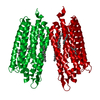
- Assembly
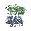
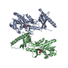
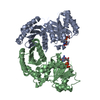
| Deposited unit | 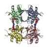
| ||||||||
|---|---|---|---|---|---|---|---|---|---|
| 1 |
| ||||||||
| 2 | 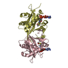
| ||||||||
| 3 | 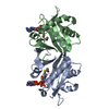
| ||||||||
| Unit cell |
|
-Components
| #1: Protein | Mass: 17922.111 Da / Num. of mol.: 4 Source method: isolated from a genetically manipulated source Source: (gene. exp.) Saccharomyces cerevisiae (brewer's yeast)Plasmid: PET13A / Production host:  Escherichia coli (E. coli) / References: UniProt: Q06592, histone acetyltransferase Escherichia coli (E. coli) / References: UniProt: Q06592, histone acetyltransferase#2: Chemical | ChemComp-ACO / Acetyl-CoA  Mass: 809.571 Da / Num. of mol.: 4 / Source method: obtained synthetically / Formula: C23H38N7O17P3S Mass: 809.571 Da / Num. of mol.: 4 / Source method: obtained synthetically / Formula: C23H38N7O17P3S#3: Water | ChemComp-HOH / | Water Mass: 18.015 Da / Num. of mol.: 190 / Source method: isolated from a natural source / Formula: H2O Mass: 18.015 Da / Num. of mol.: 190 / Source method: isolated from a natural source / Formula: H2O |
|---|
-Experimental details
-Experiment
| Experiment | Method: X-RAY DIFFRACTION / Number of used crystals: 1 |
|---|
- Sample preparation
| Crystal | Density Matthews: 3.27 Å3/Da / Density % sol: 62.4 % | |||||||||||||||||||||||||
|---|---|---|---|---|---|---|---|---|---|---|---|---|---|---|---|---|---|---|---|---|---|---|---|---|---|---|
| Crystal grow | Temperature: 277 K / Method: vapor diffusion, hanging drop / pH: 6 Details: PEG 4000, MES, CALCIUM ACETATE, pH 6.00, VAPOR DIFFUSION, HANGING DROP, temperature 277.00K | |||||||||||||||||||||||||
| Crystal | *PLUS Density % sol: 55 % | |||||||||||||||||||||||||
| Crystal grow | *PLUS Temperature: 4 ℃ / pH: 6.9 | |||||||||||||||||||||||||
| Components of the solutions | *PLUS
|
-Data collection
| Diffraction | Mean temperature: 100 K |
|---|---|
| Diffraction source | Source: SYNCHROTRON / Site: NSLS  / Beamline: X25 / Wavelength: 1.1 / Beamline: X25 / Wavelength: 1.1 |
| Detector | Type: BRANDEIS / Detector: CCD / Date: Jan 1, 1998 |
| Radiation | Protocol: SINGLE WAVELENGTH / Monochromatic (M) / Laue (L): M / Scattering type: x-ray |
| Radiation wavelength | Wavelength: 1.1 Å / Relative weight: 1 |
| Reflection | Resolution: 2.4→20 Å / Num. all: 456328 / Num. obs: 456328 / % possible obs: 100 % / Observed criterion σ(I): -3 / Redundancy: 6.5 % / Biso Wilson estimate: 28.9 Å2 / Rmerge(I) obs: 0.045 / Net I/σ(I): 21 |
| Reflection shell | Resolution: 2.4→2.53 Å / Redundancy: 8.5 % / Rmerge(I) obs: 0.113 / % possible all: 99.7 |
| Reflection | *PLUS Highest resolution: 2.4 Å / Lowest resolution: 20 Å / Num. obs: 37150 / % possible obs: 100 % / Observed criterion σ(I): -3 / Redundancy: 6.5 % / Num. measured all: 456328 |
| Reflection shell | *PLUS Redundancy: 8.5 % / Mean I/σ(I) obs: 7.66 |
- Processing
| Software |
| ||||||||||||||||||||||||||||||||||||||||||||||||||||||||||||||||||||||||||||||||
|---|---|---|---|---|---|---|---|---|---|---|---|---|---|---|---|---|---|---|---|---|---|---|---|---|---|---|---|---|---|---|---|---|---|---|---|---|---|---|---|---|---|---|---|---|---|---|---|---|---|---|---|---|---|---|---|---|---|---|---|---|---|---|---|---|---|---|---|---|---|---|---|---|---|---|---|---|---|---|---|---|---|
| Refinement | Rfactor Rfree error: 0.003 / Highest resolution: 2.4 Å / Data cutoff high absF: 10000000 / Data cutoff low absF: 0 / Isotropic thermal model: RESTRAINED / Cross valid method: THROUGHOUT / σ(F): 0 / Details: BULK SOLVENT MODEL USED
| ||||||||||||||||||||||||||||||||||||||||||||||||||||||||||||||||||||||||||||||||
| Displacement parameters | Biso mean: 22.9 Å2
| ||||||||||||||||||||||||||||||||||||||||||||||||||||||||||||||||||||||||||||||||
| Refine analyze |
| ||||||||||||||||||||||||||||||||||||||||||||||||||||||||||||||||||||||||||||||||
| Refinement step | Cycle: LAST / Highest resolution: 2.4 Å
| ||||||||||||||||||||||||||||||||||||||||||||||||||||||||||||||||||||||||||||||||
| Refine LS restraints |
| ||||||||||||||||||||||||||||||||||||||||||||||||||||||||||||||||||||||||||||||||
| LS refinement shell | Resolution: 2.4→2.55 Å / Total num. of bins used: 6 /
| ||||||||||||||||||||||||||||||||||||||||||||||||||||||||||||||||||||||||||||||||
| Software | *PLUS Name: X-PLOR / Version: 3.843 / Classification: refinement | ||||||||||||||||||||||||||||||||||||||||||||||||||||||||||||||||||||||||||||||||
| Refinement | *PLUS Lowest resolution: 20 Å / σ(F): 0 / % reflection Rfree: 9.9 % | ||||||||||||||||||||||||||||||||||||||||||||||||||||||||||||||||||||||||||||||||
| Solvent computation | *PLUS | ||||||||||||||||||||||||||||||||||||||||||||||||||||||||||||||||||||||||||||||||
| Displacement parameters | *PLUS Biso mean: 22.9 Å2 | ||||||||||||||||||||||||||||||||||||||||||||||||||||||||||||||||||||||||||||||||
| Refine LS restraints | *PLUS
| ||||||||||||||||||||||||||||||||||||||||||||||||||||||||||||||||||||||||||||||||
| LS refinement shell | *PLUS |
