 Movie
Movie Controller


+ Open data

- Basic information
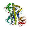
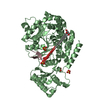
| Entry | Database: PDB / ID: 1qs1 | ||||||
|---|---|---|---|---|---|---|---|
| Title | CRYSTAL STRUCTURE OF VEGETATIVE INSECTICIDAL PROTEIN2 (VIP2) | ||||||
 Components Components | ADP-RIBOSYLTRANSFERASE Poly (ADP-ribose) polymerase Poly (ADP-ribose) polymerase | ||||||
 Keywords Keywords | TOXIN / ALPHA-BETA PROTEIN / BINARY TOXIN / INSECTICIAL PROTEIN | ||||||
| Function / homology |  Function and homology information Function and homology information | ||||||
| Biological species |  Bacillus cereus (bacteria) Bacillus cereus (bacteria) | ||||||
| Method | X-RAY DIFFRACTION / SYNCHROTRON / Resolution: 1.5 Å | ||||||
 Authors Authors | Han, S. / Craig, J.A. / Putnam, C.D. / Carozzi, N.B. / Tainer, J.A. | ||||||
 Citation Citation | Journal: Nat.Struct.Biol. / Year: 1999 Title: Evolution and mechanism from structures of an ADP-ribosylating toxin and NAD complex. Authors: Han, S. / Craig, J.A. / Putnam, C.D. / Carozzi, N.B. / Tainer, J.A. | ||||||
| History |
|
- Structure visualization
| Structure viewer | Molecule: MolmilJmol/JSmol |
|---|
- Downloads & links
-Download
| PDBx/mmCIF format | 1qs1.cif.gz | 355.3 KB | Display | PDBx/mmCIF format |
|---|---|---|---|---|
| PDB format | pdb1qs1.ent.gz | 284.3 KB | Display | PDB format |
| PDBx/mmJSON format | 1qs1.json.gz | Tree view | PDBx/mmJSON format | |
| Others |  Other downloads Other downloads |
-Validation report
| Arichive directory | https://data.pdbj.org/pub/pdb/validation_reports/qs/1qs1ftp://data.pdbj.org/pub/pdb/validation_reports/qs/1qs1 | HTTPS FTP |
|---|
-Related structure data


-Links
 PDBj
PDBj- Assembly
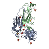
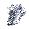
| Deposited unit | 
| ||||||||
|---|---|---|---|---|---|---|---|---|---|
| 1 | 
| ||||||||
| 2 | 
| ||||||||
| 3 | 
| ||||||||
| 4 | 
| ||||||||
| Unit cell |
|
-Components
| #1: Protein | Poly (ADP-ribose) polymerase Mass: 52519.730 Da / Num. of mol.: 4 / Fragment: MATURE VIP2 / Mutation: WILD TYPE Source method: isolated from a genetically manipulated source Source: (gene. exp.) Bacillus cereus (bacteria) / Production host: Bacillus cereus (bacteria)References: UniProt: Q844J9*PLUS, NAD+ ADP-ribosyltransferase#2: Water | ChemComp-HOH / | Water Mass: 18.015 Da / Num. of mol.: 1501 / Source method: isolated from a natural source / Formula: H2O Mass: 18.015 Da / Num. of mol.: 1501 / Source method: isolated from a natural source / Formula: H2O |
|---|
-Experimental details
-Experiment
| Experiment | Method: X-RAY DIFFRACTION / Number of used crystals: 1 |
|---|
- Sample preparation
| Crystal | Density Matthews: 2.03 Å3/Da / Density % sol: 39.28 % | ||||||||||||||||||||||||||||||
|---|---|---|---|---|---|---|---|---|---|---|---|---|---|---|---|---|---|---|---|---|---|---|---|---|---|---|---|---|---|---|---|
| Crystal grow | Temperature: 298 K / Method: vapor diffusion, hanging drop / pH: 7.5 Details: PEG200, BOG, IMIDAZOLE, MALATE, pH 7.5, VAPOR DIFFUSION, HANGING DROP, temperature 298.0K | ||||||||||||||||||||||||||||||
| Crystal grow | *PLUS Temperature: 20 ℃ / Method: vapor diffusion | ||||||||||||||||||||||||||||||
| Components of the solutions | *PLUS
|
-Data collection
| Diffraction | Mean temperature: 103 K |
|---|---|
| Diffraction source | Source: SYNCHROTRON / Site: CHESS  / Beamline: F1 / Wavelength: 1.5418 / Beamline: F1 / Wavelength: 1.5418 |
| Detector | Type: MARRESEARCH / Detector: CCD / Date: Dec 30, 1997 |
| Radiation | Protocol: SINGLE WAVELENGTH / Monochromatic (M) / Laue (L): M / Scattering type: x-ray |
| Radiation wavelength | Wavelength: 1.5418 Å / Relative weight: 1 |
| Reflection | Resolution: 1.5→20 Å / Num. all: 468627 / Num. obs: 215237 / % possible obs: 83.4 % / Observed criterion σ(I): -3 / Redundancy: 2.2 % / Biso Wilson estimate: 22.1 Å2 / Rmerge(I) obs: 0.046 / Net I/σ(I): 13 |
| Reflection shell | Resolution: 1.5→1.55 Å / Redundancy: 1.6 % / Rmerge(I) obs: 0.27 / % possible all: 69.2 |
| Reflection | *PLUS Num. measured all: 468627 |
| Reflection shell | *PLUS % possible obs: 69.2 % / Rmerge(I) obs: 0.27 |
- Processing
| Software |
| ||||||||||||||||||||||||||||||||||||||||||||||||||||||||||||
|---|---|---|---|---|---|---|---|---|---|---|---|---|---|---|---|---|---|---|---|---|---|---|---|---|---|---|---|---|---|---|---|---|---|---|---|---|---|---|---|---|---|---|---|---|---|---|---|---|---|---|---|---|---|---|---|---|---|---|---|---|---|
| Refinement | Resolution: 1.5→20 Å / σ(F): 0 / Stereochemistry target values: ENGH & HUBER
| ||||||||||||||||||||||||||||||||||||||||||||||||||||||||||||
| Displacement parameters | Biso mean: 24.77 Å2
| ||||||||||||||||||||||||||||||||||||||||||||||||||||||||||||
| Refinement step | Cycle: LAST / Resolution: 1.5→20 Å
| ||||||||||||||||||||||||||||||||||||||||||||||||||||||||||||
| Refine LS restraints |
| ||||||||||||||||||||||||||||||||||||||||||||||||||||||||||||
| LS refinement shell | Resolution: 1.5→1.57 Å / Total num. of bins used: 8
| ||||||||||||||||||||||||||||||||||||||||||||||||||||||||||||
| Software | *PLUS Name: CNS / Classification: refinement | ||||||||||||||||||||||||||||||||||||||||||||||||||||||||||||
| Refinement | *PLUS Highest resolution: 1.5 Å / Lowest resolution: 20 Å / σ(F): 0 | ||||||||||||||||||||||||||||||||||||||||||||||||||||||||||||
| Solvent computation | *PLUS | ||||||||||||||||||||||||||||||||||||||||||||||||||||||||||||
| Displacement parameters | *PLUS | ||||||||||||||||||||||||||||||||||||||||||||||||||||||||||||
| Refine LS restraints | *PLUS
| ||||||||||||||||||||||||||||||||||||||||||||||||||||||||||||
| LS refinement shell | *PLUS Highest resolution: 1.5 Å / Rfactor Rfree: 0.308 / Rfactor Rwork: 0.3 |
