 Movie
Movie Controller


+ Open data

- Basic information
| Entry | Database: PDB / ID: 1qnn | ||||||
|---|---|---|---|---|---|---|---|
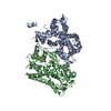
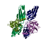
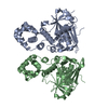
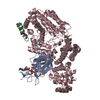
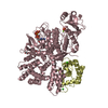
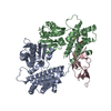
| Title | Cambialistic superoxide dismutase from Porphyromonas gingivalis | ||||||
 Components Components | SUPEROXIDE DISMUTASE | ||||||
 Keywords Keywords | OXIDOREDUCTASE | ||||||
| Function / homology |  Function and homology informationsuperoxide dismutase / superoxide dismutase activity / metal ion binding Function and homology informationsuperoxide dismutase / superoxide dismutase activity / metal ion bindingSimilarity search - Function | ||||||
| Biological species |  PORPHYROMONAS GINGIVALIS (bacteria) PORPHYROMONAS GINGIVALIS (bacteria) | ||||||
| Method | X-RAY DIFFRACTION / MOLECULAR REPLACEMENT / Resolution: 1.8 Å | ||||||
 Authors Authors | Sugio, S. / Hiraoka, B.Y. / Yamakura, F. | ||||||
 Citation Citation | Journal: Eur.J.Biochem. / Year: 2000 Title: Crystal Structure of Cambialistic Superoxide Dismutase from Porphyromonas Gingivalis Authors: Sugio, S. / Hiraoka, B.Y. / Yamakura, F. #1: Journal: Eur.J.Biochem. / Year: 1998 Title: Inactivation and Destruction of Conserved Trp159 of Fe-Superoxide Dismutase from Porphyromonas Gingivalis by Hydrogen Peroxide Authors: Yamakura, F. / Rardin, R.L. / Petsko, G.A. / Ringe, D. / Hiraoka, B.Y. / Nakayama, K. / Fujimura, T. / Taka, H. / Murayama, K. #2: Journal: FEBS Lett. / Year: 1990 Title: The Primary Structure of Superoxide Dismutase Purified from Anaerobically Maintained Bacteroides Gingivalis Authors: Amano, A. / Shizukuishi, S. / Tsunemitsu, A. / Maekawa, K. / Tsunemitsu, S. #3: Journal: J.Bacteriol. / Year: 1990 Title: Characterization of Superoxide Dismutase Purified from Either Anaerobically Maintained or Aerated Bacteroides Gingivalis Authors: Amano, A. / Shizukuishi, S. / Tamagawa, H. / Iwakura, K. / Tsunasawa, S. / Tsunemitsu, S. | ||||||
| History |
|
- Structure visualization
| Structure viewer | Molecule: MolmilJmol/JSmol |
|---|
- Downloads & links
-Download
| PDBx/mmCIF format | 1qnn.cif.gz | 167.7 KB | Display | PDBx/mmCIF format |
|---|---|---|---|---|
| PDB format | pdb1qnn.ent.gz | 134.8 KB | Display | PDB format |
| PDBx/mmJSON format | 1qnn.json.gz | Tree view | PDBx/mmJSON format | |
| Others |  Other downloads Other downloads |
-Validation report
| Arichive directory | https://data.pdbj.org/pub/pdb/validation_reports/qn/1qnnftp://data.pdbj.org/pub/pdb/validation_reports/qn/1qnn | HTTPS FTP |
|---|
-Related structure data
| Related structure data |  1isaS S: Starting model for refinement |
|---|---|
| Similar structure data |


-Links
 PDBj
PDBj
- Assembly
| Deposited unit | 
| ||||||||||||||||
|---|---|---|---|---|---|---|---|---|---|---|---|---|---|---|---|---|---|
| 1 |
| ||||||||||||||||
| Unit cell |
| ||||||||||||||||
| Noncrystallographic symmetry (NCS) | NCS oper:
|
-Components
| #1: Protein | / FE/MN-SOD Mass: 21528.133 Da / Num. of mol.: 4 Source method: isolated from a genetically manipulated source Details: EACH CHAIN CONTAINS ONE METAL ION (FE3+\:MN3+=3\:1). Source: (gene. exp.) PORPHYROMONAS GINGIVALIS (bacteria) / Plasmid: PKD210 / Gene (production host): SOD / Production host: ESCHERICHIA COLI (E. coli) / Variant (production host): QC774 / References: UniProt: P19665, superoxide dismutase#2: Chemical | ChemComp-FE / Iron  Mass: 55.845 Da / Num. of mol.: 4 / Source method: obtained synthetically / Formula: Fe Mass: 55.845 Da / Num. of mol.: 4 / Source method: obtained synthetically / Formula: Fe#3: Water | ChemComp-HOH / | Water Mass: 18.015 Da / Num. of mol.: 436 / Source method: isolated from a natural source / Formula: H2O Mass: 18.015 Da / Num. of mol.: 436 / Source method: isolated from a natural source / Formula: H2O |
|---|
-Experimental details
-Experiment
| Experiment | Method: X-RAY DIFFRACTION / Number of used crystals: 1 |
|---|
- Sample preparation
| Crystal | Density Matthews: 2.3 Å3/Da / Density % sol: 46 % | |||||||||||||||||||||||||
|---|---|---|---|---|---|---|---|---|---|---|---|---|---|---|---|---|---|---|---|---|---|---|---|---|---|---|
| Crystal grow | Temperature: 293 K / Method: vapor diffusion, hanging drop / pH: 5.8 Details: SOLUTION CONTAINING 6-9MG/ML PROTEIN AND 10MM TRIS/HCL (PH7.8) WERE USED FOR CRYSTALLIZATION. CRYSTALS WERE GROWN FROM HANGING DROPS SUSPENDED OVER A RESERVOIR SOLUTION CONTAINING 100MM ...Details: SOLUTION CONTAINING 6-9MG/ML PROTEIN AND 10MM TRIS/HCL (PH7.8) WERE USED FOR CRYSTALLIZATION. CRYSTALS WERE GROWN FROM HANGING DROPS SUSPENDED OVER A RESERVOIR SOLUTION CONTAINING 100MM POTASSIUM PHOSPHATE (PH5.8) AND 26-31% PEG4000 AT 293K WITHIN A FEW WEEKS., pH 5.80 | |||||||||||||||||||||||||
| Crystal grow | *PLUS pH: 7.8 / Method: vapor diffusion, hanging dropDetails: drop consists of equal volume of protein and reservoir solutions | |||||||||||||||||||||||||
| Components of the solutions | *PLUS
|
-Data collection
| Diffraction | Mean temperature: 293 K |
|---|---|
| Diffraction source | Source: ROTATING ANODE / Type: RIGAKU RUH2R / Wavelength: 1.5418 |
| Detector | Type: RIGAKU IMAGE PLATE / Detector: IMAGE PLATE / Date: Aug 29, 1996 / Details: YALE MIRRORS |
| Radiation | Monochromator: NI FILTER / Protocol: SINGLE WAVELENGTH / Monochromatic (M) / Laue (L): M / Scattering type: x-ray |
| Radiation wavelength | Wavelength: 1.5418 Å / Relative weight: 1 |
| Reflection | Resolution: 1.79→99.4 Å / Num. obs: 64134 / % possible obs: 88.2 % / Observed criterion σ(I): 0 / Redundancy: 5.64 % / Biso Wilson estimate: 22.1 Å2 / Rmerge(I) obs: 0.057 / Net I/σ(I): 11.6 |
| Reflection shell | Resolution: 1.79→1.86 Å / Rmerge(I) obs: 0.262 / Mean I/σ(I) obs: 2 / % possible all: 68.5 |
| Reflection shell | *PLUS % possible obs: 68.5 % |
- Processing
| Software |
| ||||||||||||||||||||||||||||||||||||||||||||||||||||||||||||||||||||||||||||||||
|---|---|---|---|---|---|---|---|---|---|---|---|---|---|---|---|---|---|---|---|---|---|---|---|---|---|---|---|---|---|---|---|---|---|---|---|---|---|---|---|---|---|---|---|---|---|---|---|---|---|---|---|---|---|---|---|---|---|---|---|---|---|---|---|---|---|---|---|---|---|---|---|---|---|---|---|---|---|---|---|---|---|
| Refinement | Method to determine structure: MOLECULAR REPLACEMENT Starting model: PDB ENTRY 1ISA Resolution: 1.8→50 Å / Rfactor Rfree error: 0.005 / Data cutoff high absF: 1000000 / Data cutoff low absF: 0.001 / Isotropic thermal model: RESTRAINED / Cross valid method: THROUGHOUT / σ(F): 0 Details: A RESOLUTION-DEPENDENT WEIGHTS AND BULK SOLVENT CORRECTION WERE APPLIED
| ||||||||||||||||||||||||||||||||||||||||||||||||||||||||||||||||||||||||||||||||
| Displacement parameters | Biso mean: 31.4 Å2 | ||||||||||||||||||||||||||||||||||||||||||||||||||||||||||||||||||||||||||||||||
| Refine analyze |
| ||||||||||||||||||||||||||||||||||||||||||||||||||||||||||||||||||||||||||||||||
| Refinement step | Cycle: LAST / Resolution: 1.8→50 Å
| ||||||||||||||||||||||||||||||||||||||||||||||||||||||||||||||||||||||||||||||||
| Refine LS restraints |
| ||||||||||||||||||||||||||||||||||||||||||||||||||||||||||||||||||||||||||||||||
| LS refinement shell | Resolution: 1.8→1.88 Å / Rfactor Rfree error: 0.025 / Total num. of bins used: 8
| ||||||||||||||||||||||||||||||||||||||||||||||||||||||||||||||||||||||||||||||||
| Xplor file |
| ||||||||||||||||||||||||||||||||||||||||||||||||||||||||||||||||||||||||||||||||
| Software | *PLUS Name: X-PLOR / Version: 3.851 / Classification: refinement | ||||||||||||||||||||||||||||||||||||||||||||||||||||||||||||||||||||||||||||||||
| Refine LS restraints | *PLUS
|
