 Movie
Movie Controller

[English] 日本語
 Yorodumi
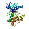
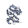
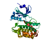
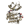
- PDB-1ql6: THE CATALYTIC MECHANISM OF PHOSPHORYLASE KINASE PROBED BY MUTATIO... -
+ Open data

- Basic information
| Entry | Database: PDB / ID: 1ql6 | ||||||
|---|---|---|---|---|---|---|---|
| Title | THE CATALYTIC MECHANISM OF PHOSPHORYLASE KINASE PROBED BY MUTATIONAL STUDIES | ||||||
 Components Components | PHOSPHORYLASE KINASE | ||||||
 Keywords Keywords | KINASE (GLYCOGEN METABOLISM) / GLYCOGEN METABOLISM / TRANSFERASE / SERINE/THREONINE-PROTEIN / KINASE / ATP-BINDING / CALMODULIN-BINDING | ||||||
| Function / homology |  Function and homology informationphosphorylase kinase / phosphorylase kinase activity / phosphorylase kinase complex / tau-protein kinase / glycogen biosynthetic process / tau-protein kinase activity / skeletal muscle myofibril / calmodulin binding / non-specific serine/threonine protein kinase / phosphorylation ...phosphorylase kinase / phosphorylase kinase activity / phosphorylase kinase complex / tau-protein kinase / glycogen biosynthetic process / tau-protein kinase activity / skeletal muscle myofibril / calmodulin binding / non-specific serine/threonine protein kinase / phosphorylation / protein serine kinase activity / ATP binding Function and homology informationphosphorylase kinase / phosphorylase kinase activity / phosphorylase kinase complex / tau-protein kinase / glycogen biosynthetic process / tau-protein kinase activity / skeletal muscle myofibril / calmodulin binding / non-specific serine/threonine protein kinase / phosphorylation ...phosphorylase kinase / phosphorylase kinase activity / phosphorylase kinase complex / tau-protein kinase / glycogen biosynthetic process / tau-protein kinase activity / skeletal muscle myofibril / calmodulin binding / non-specific serine/threonine protein kinase / phosphorylation / protein serine kinase activity / ATP bindingSimilarity search - Function | ||||||
| Biological species |  ORYCTOLAGUS CUNICULUS (rabbit) ORYCTOLAGUS CUNICULUS (rabbit) | ||||||
| Method | X-RAY DIFFRACTION / SYNCHROTRON / MOLECULAR REPLACEMENT / Resolution: 2.4 Å | ||||||
 Authors Authors | Skamnaki, V.T. / Owen, D.J. / Noble, M.E.M. / Lowe, E.D. / Oikonomakos, N.G. / Johnson, L.N. | ||||||
 Citation Citation | Journal: Biochemistry / Year: 1999 Title: Catalytic Mechanism of Phosphorylase Kinase Probed by Mutational Studies. Authors: Skamnaki, V.T. / Owen, D.J. / Noble, M.E. / Lowe, E.D. / Lowe, G. / Oikonomakos, N.G. / Johnson, L.N. #1: Journal: Embo J. / Year: 1997Title: The Crystal Structure of a Phosphorylase Kinase Peptide Substrate Complex: Kinase Substrate Recognition Authors: Lowe, E.D. / Noble, M.E.M. / Skamnaki, V.T. / Oikonomakos, N.G. / Owen, D.J. / Johnson, L.N. | ||||||
| History |
| ||||||
| Remark 650 | HELIX DETERMINATION METHOD: AUTHOR PROVIDED. | ||||||
| Remark 700 | SHEET DETERMINATION METHOD: AUTHOR PROVIDED. |
- Structure visualization
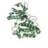
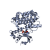
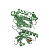
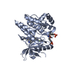
| Structure viewer | Molecule: MolmilJmol/JSmol |
|---|
- Downloads & links
-Download
| PDBx/mmCIF format | 1ql6.cif.gz | 76 KB | Display | PDBx/mmCIF format |
|---|---|---|---|---|
| PDB format | pdb1ql6.ent.gz | 54.5 KB | Display | PDB format |
| PDBx/mmJSON format | 1ql6.json.gz | Tree view | PDBx/mmJSON format | |
| Others |  Other downloads Other downloads |
-Validation report
| Arichive directory | https://data.pdbj.org/pub/pdb/validation_reports/ql/1ql6ftp://data.pdbj.org/pub/pdb/validation_reports/ql/1ql6 | HTTPS FTP |
|---|
-Related structure data
| Related structure data | 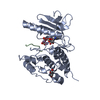 2phkS S: Starting model for refinement |
|---|---|
| Similar structure data |
-Links
 PDBj
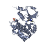
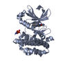
PDBj- Assembly
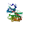
| Deposited unit | 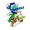
| ||||||||
|---|---|---|---|---|---|---|---|---|---|
| 1 | 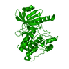
| ||||||||
| Unit cell |
|
-Components
| #1: Protein | / RABBIT MUSCLE PHOSPHORYLASE KINASE Mass: 34274.262 Da / Num. of mol.: 1 / Fragment: GAMMA SUBUNIT, TRUNCATED TO RESIDUES 1 - 298 / Mutation: YES Source method: isolated from a genetically manipulated source Details: LIGANDS ATP AND MN(II) / Source: (gene. exp.) ORYCTOLAGUS CUNICULUS (rabbit) / Tissue: SKELETAL MUSCLE / Plasmid: PMW172 / Production host:  ESCHERICHIA COLI (E. coli) / References: UniProt: P00518, EC: 2.7.1.38 ESCHERICHIA COLI (E. coli) / References: UniProt: P00518, EC: 2.7.1.38 | ||||
|---|---|---|---|---|---|
| #2: Chemical | ChemComp-ATP / Adenosine triphosphate  Mass: 507.181 Da / Num. of mol.: 1 / Source method: obtained synthetically / Formula: C10H16N5O13P3 / Comment: ATP, energy-carrying molecule*YM Mass: 507.181 Da / Num. of mol.: 1 / Source method: obtained synthetically / Formula: C10H16N5O13P3 / Comment: ATP, energy-carrying molecule*YM | ||||
| #3: Chemical |   Mass: 54.938 Da / Num. of mol.: 2 / Source method: obtained synthetically / Formula: Mn Mass: 54.938 Da / Num. of mol.: 2 / Source method: obtained synthetically / Formula: Mn#4: Chemical | ChemComp-SO4 / | Sulfate  Mass: 96.063 Da / Num. of mol.: 1 / Source method: obtained synthetically / Formula: SO4 Mass: 96.063 Da / Num. of mol.: 1 / Source method: obtained synthetically / Formula: SO4#5: Water | ChemComp-HOH / | Water Mass: 18.015 Da / Num. of mol.: 104 / Source method: isolated from a natural source / Formula: H2O Mass: 18.015 Da / Num. of mol.: 104 / Source method: isolated from a natural source / Formula: H2O |
-Experimental details
-Experiment
| Experiment | Method: X-RAY DIFFRACTION / Number of used crystals: 1 |
|---|
- Sample preparation
| Crystal | Density Matthews: 2.66 Å3/Da / Density % sol: 50 % | ||||||||||||||||||||||||||||||||||||||||||||||||||||||||||||||||||||||||||||||||||||||||||
|---|---|---|---|---|---|---|---|---|---|---|---|---|---|---|---|---|---|---|---|---|---|---|---|---|---|---|---|---|---|---|---|---|---|---|---|---|---|---|---|---|---|---|---|---|---|---|---|---|---|---|---|---|---|---|---|---|---|---|---|---|---|---|---|---|---|---|---|---|---|---|---|---|---|---|---|---|---|---|---|---|---|---|---|---|---|---|---|---|---|---|---|
| Crystal grow | pH: 6.9 / Details: pH 6.90 | ||||||||||||||||||||||||||||||||||||||||||||||||||||||||||||||||||||||||||||||||||||||||||
| Crystal grow | *PLUS Temperature: 14 ℃ / pH: 8.2 / Method: vapor diffusion, hanging drop | ||||||||||||||||||||||||||||||||||||||||||||||||||||||||||||||||||||||||||||||||||||||||||
| Components of the solutions | *PLUS
|
-Data collection
| Diffraction | Mean temperature: 100 K |
|---|---|
| Diffraction source | Source: SYNCHROTRON / Site: ESRF  / Type: ESRF / Wavelength: 0.993 / Type: ESRF / Wavelength: 0.993 |
| Detector | Type: MARRESEARCH / Detector: IMAGE PLATE / Date: Nov 7, 1995 |
| Radiation | Protocol: SINGLE WAVELENGTH / Monochromatic (M) / Laue (L): M / Scattering type: x-ray |
| Radiation wavelength | Wavelength: 0.993 Å / Relative weight: 1 |
| Reflection | Resolution: 2.4→20 Å / Num. obs: 12745 / % possible obs: 86.6 % / Observed criterion σ(I): 2 / Redundancy: 3.2 % / Rmerge(I) obs: 0.064 / Rsym value: 0.048 / Net I/σ(I): 10.1 |
| Reflection | *PLUS Num. measured all: 40795 / Rmerge(I) obs: 0.048 |
| Reflection shell | *PLUS % possible obs: 78.5 % / Rmerge(I) obs: 0.221 |
- Processing
| Software |
| ||||||||||||||||||||||||||||||||||||||||||||||||||||||||||||||||||||||||||||||||||||
|---|---|---|---|---|---|---|---|---|---|---|---|---|---|---|---|---|---|---|---|---|---|---|---|---|---|---|---|---|---|---|---|---|---|---|---|---|---|---|---|---|---|---|---|---|---|---|---|---|---|---|---|---|---|---|---|---|---|---|---|---|---|---|---|---|---|---|---|---|---|---|---|---|---|---|---|---|---|---|---|---|---|---|---|---|---|
| Refinement | Method to determine structure: MOLECULAR REPLACEMENT Starting model: PDB ENTRY 2PHK Resolution: 2.4→20 Å / Cross valid method: THROUGHOUT / σ(F): 2
| ||||||||||||||||||||||||||||||||||||||||||||||||||||||||||||||||||||||||||||||||||||
| Refinement step | Cycle: LAST / Resolution: 2.4→20 Å
| ||||||||||||||||||||||||||||||||||||||||||||||||||||||||||||||||||||||||||||||||||||
| Refine LS restraints |
|
