 Movie
Movie Controller

[English] 日本語
 Yorodumi
- PDB-1ql4: Structure of the soluble domain of cytochrome c552 from Paracoccu... -
+ Open data

- Basic information
| Entry | Database: PDB / ID: 1ql4 | ||||||
|---|---|---|---|---|---|---|---|
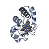
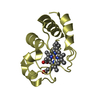
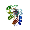
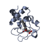
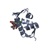
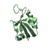
| Title | Structure of the soluble domain of cytochrome c552 from Paracoccus denitrificans in the oxidised state | ||||||
 Components Components | CYTOCHROME C552 | ||||||
 Keywords Keywords |  ELECTRON TRANSPORT PROTEIN (CYTOCHROME) / ELECTRON TRANSFER / OXIDISED ELECTRON TRANSPORT PROTEIN (CYTOCHROME) / ELECTRON TRANSFER / OXIDISED | ||||||
| Function / homology |  Function and homology informationelectron transfer activity / heme binding / metal ion binding / plasma membrane Function and homology informationelectron transfer activity / heme binding / metal ion binding / plasma membraneSimilarity search - Function | ||||||
| Biological species |  PARACOCCUS DENITRIFICANS (bacteria) PARACOCCUS DENITRIFICANS (bacteria) | ||||||
| Method | X-RAY DIFFRACTION / SYNCHROTRON / MAD / Resolution: 1.5 Å | ||||||
 Authors Authors | Harrenga, A. / Reincke, B. / Rueterjans, H. / Ludwig, B. / Michel, H. | ||||||
 Citation Citation | Journal: J.Mol.Biol. / Year: 2000 Title: Structure of the Soluble Domain of Cytochrome C552 from Paracoccus Denitrificans in the Oxidized and Reduced States Authors: Harrenga, A. / Reincke, B. / Rueterjans, H. / Ludwig, B. / Michel, H. | ||||||
| History |
|
- Structure visualization
| Structure viewer | Molecule: MolmilJmol/JSmol |
|---|
- Downloads & links
-Download
| PDBx/mmCIF format | 1ql4.cif.gz | 94.4 KB | Display | PDBx/mmCIF format |
|---|---|---|---|---|
| PDB format | pdb1ql4.ent.gz | 79.3 KB | Display | PDB format |
| PDBx/mmJSON format | 1ql4.json.gz | Tree view | PDBx/mmJSON format | |
| Others |  Other downloads Other downloads |
-Validation report
| Arichive directory | https://data.pdbj.org/pub/pdb/validation_reports/ql/1ql4ftp://data.pdbj.org/pub/pdb/validation_reports/ql/1ql4 | HTTPS FTP |
|---|
-Related structure data


-Links
 PDBj
PDBj


- Assembly
| Deposited unit | 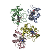
| ||||||||
|---|---|---|---|---|---|---|---|---|---|
| 1 | 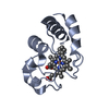
| ||||||||
| 2 | 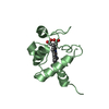
| ||||||||
| 3 | 
| ||||||||
| 4 | 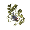
| ||||||||
| Unit cell |
|
-Components
| #1: Protein | Mass: 10422.782 Da / Num. of mol.: 4 / Fragment: SOLUBLE DOMAIN Source method: isolated from a genetically manipulated source Source: (gene. exp.) PARACOCCUS DENITRIFICANS (bacteria) / Cellular location: MEMBRANE-BOUNDBiological membrane / Production host: ESCHERICHIA COLI (E. coli) / References: UniProt: P54820#2: Chemical | ChemComp-HEC / Heme C  Mass: 618.503 Da / Num. of mol.: 4 / Source method: obtained synthetically / Formula: C34H34FeN4O4 Mass: 618.503 Da / Num. of mol.: 4 / Source method: obtained synthetically / Formula: C34H34FeN4O4#3: Water | ChemComp-HOH / | Water Mass: 18.015 Da / Num. of mol.: 550 / Source method: isolated from a natural source / Formula: H2O Mass: 18.015 Da / Num. of mol.: 550 / Source method: isolated from a natural source / Formula: H2O |
|---|
-Experimental details
-Experiment
| Experiment | Method: X-RAY DIFFRACTION / Number of used crystals: 1 |
|---|
- Sample preparation
| Crystal | Density Matthews: 2.47 Å3/Da / Density % sol: 49.7 % | ||||||||||||||||||||||||||||||||||||||||||
|---|---|---|---|---|---|---|---|---|---|---|---|---|---|---|---|---|---|---|---|---|---|---|---|---|---|---|---|---|---|---|---|---|---|---|---|---|---|---|---|---|---|---|---|
| Crystal grow | pH: 4.5 / Details: pH 4.50 | ||||||||||||||||||||||||||||||||||||||||||
| Crystal grow | *PLUS Temperature: 18 ℃ / pH: 7.2 / Method: vapor diffusion, hanging drop | ||||||||||||||||||||||||||||||||||||||||||
| Components of the solutions | *PLUS
|
-Data collection
| Diffraction | Mean temperature: 100 K | |||||||||
|---|---|---|---|---|---|---|---|---|---|---|
| Diffraction source | Source: SYNCHROTRON / Site: EMBL/DESY, HAMBURG  / Beamline: BW7A / Wavelength: 1.0972, 1.7370 / Beamline: BW7A / Wavelength: 1.0972, 1.7370 | |||||||||
| Radiation | Protocol: MAD / Monochromatic (M) / Laue (L): M / Scattering type: x-ray | |||||||||
| Radiation wavelength |
| |||||||||
| Reflection | Resolution: 1.5→15 Å / Num. obs: 67817 / % possible obs: 99.9 % / Redundancy: 3.5 % / Biso Wilson estimate: 15 Å2 / Rmerge(I) obs: 0.056 / Net I/σ(I): 20.2 | |||||||||
| Reflection shell | Resolution: 1.5→1.53 Å / Redundancy: 3.1 % / Rmerge(I) obs: 0.218 / Mean I/σ(I) obs: 4.2 / % possible all: 99.9 | |||||||||
| Reflection | *PLUS Highest resolution: 1.5 Å / Lowest resolution: 15 Å / Redundancy: 3.5 % / Num. measured all: 241918 / Rmerge(I) obs: 0.056 | |||||||||
| Reflection shell | *PLUS % possible obs: 99.9 % / Redundancy: 3.1 % / Rmerge(I) obs: 0.218 / Mean I/σ(I) obs: 4.2 |
- Processing
| Software |
| ||||||||||||||||||||||||||||||||||||||||||||||||||||||||||||||||||||||||||||||||
|---|---|---|---|---|---|---|---|---|---|---|---|---|---|---|---|---|---|---|---|---|---|---|---|---|---|---|---|---|---|---|---|---|---|---|---|---|---|---|---|---|---|---|---|---|---|---|---|---|---|---|---|---|---|---|---|---|---|---|---|---|---|---|---|---|---|---|---|---|---|---|---|---|---|---|---|---|---|---|---|---|---|
| Refinement | Method to determine structure: MAD / Resolution: 1.5→15 Å / Rfactor Rfree error: 0.003 / Data cutoff high absF: 0 / Isotropic thermal model: RESTRAINED / Cross valid method: THROUGHOUT / σ(F): 0
| ||||||||||||||||||||||||||||||||||||||||||||||||||||||||||||||||||||||||||||||||
| Solvent computation | Solvent model: USED | ||||||||||||||||||||||||||||||||||||||||||||||||||||||||||||||||||||||||||||||||
| Displacement parameters | Biso mean: 17.5 Å2
| ||||||||||||||||||||||||||||||||||||||||||||||||||||||||||||||||||||||||||||||||
| Refine analyze |
| ||||||||||||||||||||||||||||||||||||||||||||||||||||||||||||||||||||||||||||||||
| Refinement step | Cycle: LAST / Resolution: 1.5→15 Å
| ||||||||||||||||||||||||||||||||||||||||||||||||||||||||||||||||||||||||||||||||
| Refine LS restraints |
| ||||||||||||||||||||||||||||||||||||||||||||||||||||||||||||||||||||||||||||||||
| LS refinement shell | Resolution: 1.5→1.59 Å / Rfactor Rfree error: 0.01 / Total num. of bins used: 6
| ||||||||||||||||||||||||||||||||||||||||||||||||||||||||||||||||||||||||||||||||
| Software | *PLUS Name: CNS / Version: 0.3 / Classification: refinement | ||||||||||||||||||||||||||||||||||||||||||||||||||||||||||||||||||||||||||||||||
| Refine LS restraints | *PLUS
|
