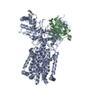
 Movie
Movie Controller


+ Open data

- Basic information
| Entry | Database: PDB / ID: 1qhm | ||||||
|---|---|---|---|---|---|---|---|
| Title | ESCHERICHIA COLI PYRUVATE FORMATE LYASE LARGE DOMAIN | ||||||
 Components Components | PYRUVATE FORMATE-LYASE | ||||||
 Keywords Keywords | LYASE/TRANSFERASE /  PYRUVATE FORMATE LYASE / ANAEROBIC / HOMODIMER / ENZYME MECHANISM / LYASE-TRANSFERASE COMPLEX PYRUVATE FORMATE LYASE / ANAEROBIC / HOMODIMER / ENZYME MECHANISM / LYASE-TRANSFERASE COMPLEX | ||||||
| Function / homology |  Function and homology informationformate C-acetyltransferase / formate C-acetyltransferase activity / glucose metabolic process / membrane / cytosol Function and homology informationformate C-acetyltransferase / formate C-acetyltransferase activity / glucose metabolic process / membrane / cytosolSimilarity search - Function | ||||||
| Biological species |  Escherichia coli (E. coli) Escherichia coli (E. coli) | ||||||
| Method | X-RAY DIFFRACTION / SYNCHROTRON / MIRAS / Resolution: 2.8 Å | ||||||
 Authors Authors | Leppanen, V.-M. / Merckel, M.C. / Ollis, D.L. / Wong, K.K. / Kozarich, J.W. / Goldman, A. | ||||||
 Citation Citation | Journal: Structure Fold.Des. / Year: 1999 Title: Pyruvate formate lyase is structurally homologous to type I ribonucleotide reductase. Authors: Leppanen, V.M. / Merckel, M.C. / Ollis, D.L. / Wong, K.K. / Kozarich, J.W. / Goldman, A. #1: Journal: Acta Crystallogr.,Sect.D / Year: 1999Title: Purification and Crystallization of a Proteolytic Fragment of Escherichia coli Pyruvate Formate-Lyase Authors: Leppanen, V.-M. / Parast, C.V. / Wong, K.K. / Kozarich, J.W. / Goldman, A. | ||||||
| History |
|
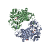
- Structure visualization
| Structure viewer | Molecule: MolmilJmol/JSmol |
|---|
- Downloads & links
-Download
| PDBx/mmCIF format | 1qhm.cif.gz | 228.2 KB | Display | PDBx/mmCIF format |
|---|---|---|---|---|
| PDB format | pdb1qhm.ent.gz | 185.3 KB | Display | PDB format |
| PDBx/mmJSON format | 1qhm.json.gz | Tree view | PDBx/mmJSON format | |
| Others |  Other downloads Other downloads |
-Validation report
| Arichive directory | https://data.pdbj.org/pub/pdb/validation_reports/qh/1qhmftp://data.pdbj.org/pub/pdb/validation_reports/qh/1qhm | HTTPS FTP |
|---|
-Related structure data
| Similar structure data |
|---|


-Links
 PDBj
PDBj- Assembly
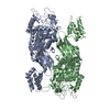
| Deposited unit | 
| ||||||||
|---|---|---|---|---|---|---|---|---|---|
| 1 |
| ||||||||
| Unit cell |
| ||||||||
| Noncrystallographic symmetry (NCS) | NCS oper: (Code: given Matrix: (-0.991906, -0.126812, -0.006467), Vector : |
-Components
| #1: Protein | Mass: 70539.180 Da / Num. of mol.: 2 / Fragment: RESIDUES 1-624 Source method: isolated from a genetically manipulated source Source: (gene. exp.) Escherichia coli (E. coli) / Cellular location: CYTOPLASM / Gene: PFL / Plasmid: PKK-TRYPFL / Species (production host): Escherichia coli / Gene (production host): PFL / Production host: Escherichia coli BL21(DE3) (bacteria) / Strain (production host): BL21(DE3) / References: UniProt: P09373, formate C-acetyltransferase#2: Water | ChemComp-HOH / | Water Mass: 18.015 Da / Num. of mol.: 234 / Source method: isolated from a natural source / Formula: H2O Mass: 18.015 Da / Num. of mol.: 234 / Source method: isolated from a natural source / Formula: H2O |
|---|
-Experimental details
-Experiment
| Experiment | Method: X-RAY DIFFRACTION / Number of used crystals: 1 |
|---|
- Sample preparation
| Crystal | Density Matthews: 4.4 Å3/Da / Density % sol: 72 % Description: IN ADDITION TO THE MIRAS DATA, A SEMET SUBSTITUENT AND MERCURY MAD DATA WERE COLLECTED ON THE BEAMLINE BW7A AT EMBL-HAMBURG OUTSTATION | ||||||||||||||||||||||||
|---|---|---|---|---|---|---|---|---|---|---|---|---|---|---|---|---|---|---|---|---|---|---|---|---|---|
| Crystal grow | pH: 7.6 Details: 18% PEG-1000, 0.1 M HEPES/HCL PH7.6, VAPOUR DIFFUSION | ||||||||||||||||||||||||
| Crystal grow | *PLUS Temperature: 294 K / Method: vapor diffusion, sitting dropDetails: Leppanen, V.-M., (1999) Acta Crystallogr., Sect.D, 55, 531. | ||||||||||||||||||||||||
| Components of the solutions | *PLUS
|
-Data collection
| Diffraction | Mean temperature: 100 K |
|---|---|
| Diffraction source | Source: SYNCHROTRON / Site: ESRF  / Beamline: ID14-3 / Wavelength: 0.933 / Beamline: ID14-3 / Wavelength: 0.933 |
| Detector | Detector: CCD / Date: May 1, 1999 |
| Radiation | Protocol: SINGLE WAVELENGTH / Monochromatic (M) / Laue (L): M / Scattering type: x-ray |
| Radiation wavelength | Wavelength: 0.933 Å / Relative weight: 1 |
| Reflection | Resolution: 2.8→20 Å / Num. obs: 59411 / % possible obs: 99.4 % / Redundancy: 5.3 % / Biso Wilson estimate: 56.6 Å2 / Rsym value: 5.2 / Net I/σ(I): 22.5 |
| Reflection shell | Resolution: 2.8→2.86 Å / Mean I/σ(I) obs: 4.7 / Rsym value: 43.7 / % possible all: 99.2 |
| Reflection | *PLUS Rmerge(I) obs: 0.045 |
| Reflection shell | *PLUS % possible obs: 96.7 % / Rmerge(I) obs: 0.195 / Mean I/σ(I) obs: 3.9 |
- Processing
| Software |
| ||||||||||||||||||||||||||||||||||||||||||||||||||||||||||||
|---|---|---|---|---|---|---|---|---|---|---|---|---|---|---|---|---|---|---|---|---|---|---|---|---|---|---|---|---|---|---|---|---|---|---|---|---|---|---|---|---|---|---|---|---|---|---|---|---|---|---|---|---|---|---|---|---|---|---|---|---|---|
| Refinement | Method to determine structure: MIRAS / Resolution: 2.8→20 Å / Rfactor Rfree error: 0.003 / Data cutoff high rms absF: 2705139.55 / Isotropic thermal model: GROUP / Cross valid method: THROUGHOUT / σ(F): 0
| ||||||||||||||||||||||||||||||||||||||||||||||||||||||||||||
| Solvent computation | Solvent model: FLAT MODEL / Bsol: 40 Å2 / ksol: 0.3 e/Å3 | ||||||||||||||||||||||||||||||||||||||||||||||||||||||||||||
| Displacement parameters | Biso mean: 63.4 Å2
| ||||||||||||||||||||||||||||||||||||||||||||||||||||||||||||
| Refine analyze |
| ||||||||||||||||||||||||||||||||||||||||||||||||||||||||||||
| Refinement step | Cycle: LAST / Resolution: 2.8→20 Å
| ||||||||||||||||||||||||||||||||||||||||||||||||||||||||||||
| Refine LS restraints |
| ||||||||||||||||||||||||||||||||||||||||||||||||||||||||||||
| LS refinement shell | Resolution: 2.8→2.88 Å / Rfactor Rfree error: 0.021 / Total num. of bins used: 12
| ||||||||||||||||||||||||||||||||||||||||||||||||||||||||||||
| Xplor file |
| ||||||||||||||||||||||||||||||||||||||||||||||||||||||||||||
| Software | *PLUS Name: CNS / Version: 0.4 / Classification: refinement | ||||||||||||||||||||||||||||||||||||||||||||||||||||||||||||
| Refinement | *PLUS σ(F): 0 / % reflection Rfree: 9.1 % / Rfactor obs: 0.228 | ||||||||||||||||||||||||||||||||||||||||||||||||||||||||||||
| Solvent computation | *PLUS | ||||||||||||||||||||||||||||||||||||||||||||||||||||||||||||
| Displacement parameters | *PLUS Biso mean: 63.4 Å2 | ||||||||||||||||||||||||||||||||||||||||||||||||||||||||||||
| Refine LS restraints | *PLUS
| ||||||||||||||||||||||||||||||||||||||||||||||||||||||||||||
| LS refinement shell | *PLUS Rfactor Rfree: 0.408 / % reflection Rfree: 8 % / Rfactor Rwork: 0.376 |
