regulation of DNA binding / adiponectin-activated signaling pathway / positive regulation of activin receptor signaling pathway / UTP-C complex / endothelial tube morphogenesis / negative regulation of viral life cycle / regulation of transcription by RNA polymerase III / protein kinase regulator activity / WNT mediated activation of DVL / Condensation of Prometaphase Chromosomes ...regulation of DNA binding / adiponectin-activated signaling pathway / positive regulation of activin receptor signaling pathway / UTP-C complex / endothelial tube morphogenesis / negative regulation of viral life cycle / regulation of transcription by RNA polymerase III / protein kinase regulator activity / WNT mediated activation of DVL / Condensation of Prometaphase Chromosomes / protein kinase CK2 complex / symbiont-mediated disruption of host cell PML body / Maturation of hRSV A proteins / Receptor Mediated Mitophagy / Synthesis of PC / RUNX1 interacts with co-factors whose precise effect on RUNX1 targets is not known / positive regulation of SMAD protein signal transduction / negative regulation of blood vessel endothelial cell migration / Signal transduction by L1 / peptidyl-threonine phosphorylation / PML body / Wnt signaling pathway / Regulation of PTEN stability and activity / Cooperation of PDCL (PhLP1) and TRiC/CCT in G-protein beta folding / KEAP1-NFE2L2 pathway / protein-macromolecule adaptor activity / protein-containing complex assembly / secretory granule lumen / Regulation of TP53 Activity through Phosphorylation / RNA polymerase II-specific DNA-binding transcription factor binding / ficolin-1-rich granule lumen / protein domain specific binding / negative regulation of cell population proliferation / signaling receptor binding / protein serine/threonine kinase activity / chromatin binding / chromatin / Neutrophil degranulation / signal transduction / extracellular exosome / extracellular region / nucleoplasm / identical protein binding / metal ion binding / nucleus / cytosol / cytoplasm Similarity search - Function
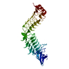
protein kinase ck2 holoenzyme, chain C, domain 1 / protein kinase ck2 holoenzyme, chain C, domain 1 / Casein kinase II, regulatory subunit / Casein kinase II, regulatory subunit, N-terminal / Casein kinase II subunit beta-like / Casein kinase II regulatory subunit / Casein kinase II regulatory subunit signature. / Casein kinase II regulatory subunit / N-terminal domain of TfIIb - #20 / N-terminal domain of TfIIb ...protein kinase ck2 holoenzyme, chain C, domain 1 / protein kinase ck2 holoenzyme, chain C, domain 1 / Casein kinase II, regulatory subunit / Casein kinase II, regulatory subunit, N-terminal / Casein kinase II subunit beta-like / Casein kinase II regulatory subunit / Casein kinase II regulatory subunit signature. / Casein kinase II regulatory subunit / N-terminal domain of TfIIb - #20 / N-terminal domain of TfIIb / Single Sheet / Orthogonal Bundle / Mainly Beta / Mainly Alpha Similarity search - Domain/homology
Mass: 18.015 Da / Num. of mol.: 324 / Source method: isolated from a natural source / Formula: H2O
Nonpolymer details
ZINC FINGER
Sequence details
RESIDUES 183-215 HAVE BEEN DELETED
-
Experimental details
-
Experiment
Experiment
Method: X-RAY DIFFRACTION / Number of used crystals: 1
-
Sample preparation
Crystal
Density Matthews: 2.7 Å3/Da / Density % sol: 60 %
Crystal grow
Temperature: 281 K / Method: vapor diffusion, sitting drop / pH: 9 Details: CRYSTALS OF CK2 BETA (1-182) WERE GROWN AT 281 K IN 4 MICROLITER SITTING DROPS CONTAINING A 1:1 MIXTURE OF PROTEIN SOLUTION (18 MG/ML) IN 10 MM TRIS (PH 7) AND RESERVOIR SOLUTION WITH 15% ...Details: CRYSTALS OF CK2 BETA (1-182) WERE GROWN AT 281 K IN 4 MICROLITER SITTING DROPS CONTAINING A 1:1 MIXTURE OF PROTEIN SOLUTION (18 MG/ML) IN 10 MM TRIS (PH 7) AND RESERVOIR SOLUTION WITH 15% PEG 5K MME, 500 MM NACL, 460 MM MGCL2 3% DIOXANE, pH 9, VAPOR DIFFUSION, SITTING DROP
In the structure databanks used in Yorodumi, some data are registered as the other names, "COVID-19 virus" and "2019-nCoV". Here are the details of the virus and the list of structure data.
Jan 31, 2019. EMDB accession codes are about to change! (news from PDBe EMDB page)
EMDB accession codes are about to change! (news from PDBe EMDB page)
The allocation of 4 digits for EMDB accession codes will soon come to an end. Whilst these codes will remain in use, new EMDB accession codes will include an additional digit and will expand incrementally as the available range of codes is exhausted. The current 4-digit format prefixed with “EMD-” (i.e. EMD-XXXX) will advance to a 5-digit format (i.e. EMD-XXXXX), and so on. It is currently estimated that the 4-digit codes will be depleted around Spring 2019, at which point the 5-digit format will come into force.
The EM Navigator/Yorodumi systems omit the EMD- prefix.
Related info.:Q: What is EMD? / ID/Accession-code notation in Yorodumi/EM Navigator
Yorodumi is a browser for structure data from EMDB, PDB, SASBDB, etc.
This page is also the successor to EM Navigator detail page, and also detail information page/front-end page for Omokage search.
The word "yorodu" (or yorozu) is an old Japanese word meaning "ten thousand". "mi" (miru) is to see.
Related info.:EMDB / PDB / SASBDB / Comparison of 3 databanks / Yorodumi Search / Aug 31, 2016. New EM Navigator & Yorodumi / Yorodumi Papers / Jmol/JSmol / Function and homology information / Changes in new EM Navigator and Yorodumi
 Movie
Movie Controller

 Yorodumi
 Open data
 Basic information
 Components
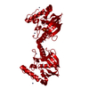
 Casein kinase 2
  Keywords
 Function and homology information
 Authors
 Citation
 Structure visualization
 Downloads & links
 Other downloads
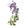


 PDBj
PDBj


 Assembly
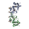


 Mass: 65.409 Da / Num. of mol.: 2 / Source method: obtained synthetically / Formula: Zn
 Mass: 24.305 Da / Num. of mol.: 1 / Source method: obtained synthetically / Formula: Mg
 Mass: 18.015 Da / Num. of mol.: 324 / Source method: isolated from a natural source / Formula: H2O
 Sample preparation
 / Beamline: BM14 / Wavelength: 0.88, 0.978668, 0.978418
 Processing