 Movie
Movie Controller


+ Open data

- Basic information
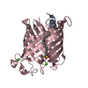
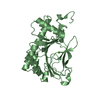
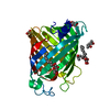
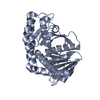
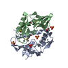
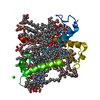
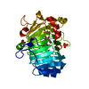
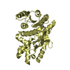
| Entry | Database: PDB / ID: 1qd5 | ||||||
|---|---|---|---|---|---|---|---|
| Title | OUTER MEMBRANE PHOSPHOLIPASE A FROM ESCHERICHIA COLI | ||||||
 Components Components | OUTER MEMBRANE PHOSPHOLIPASE A | ||||||
 Keywords Keywords |  MEMBRANE PROTEIN / ANTI-PARALLEL BETA BARREL / membrane phospholipase MEMBRANE PROTEIN / ANTI-PARALLEL BETA BARREL / membrane phospholipase | ||||||
| Function / homology |  Function and homology informationphospholipase A1 / phosphatidylserine 1-acylhydrolase activity / 1-acyl-2-lysophosphatidylserine acylhydrolase activity / phospholipase A1 activity / phospholipase activity / phosphatidylglycerol metabolic process / lysophospholipase activity / phospholipase A2 activity / phospholipase A2 / lipid catabolic process ...phospholipase A1 / phosphatidylserine 1-acylhydrolase activity / 1-acyl-2-lysophosphatidylserine acylhydrolase activity / phospholipase A1 activity / phospholipase activity / phosphatidylglycerol metabolic process / lysophospholipase activity / phospholipase A2 activity / phospholipase A2 / lipid catabolic process / cell outer membrane / calcium ion binding / protein homodimerization activity Function and homology informationphospholipase A1 / phosphatidylserine 1-acylhydrolase activity / 1-acyl-2-lysophosphatidylserine acylhydrolase activity / phospholipase A1 activity / phospholipase activity / phosphatidylglycerol metabolic process / lysophospholipase activity / phospholipase A2 activity / phospholipase A2 / lipid catabolic process ...phospholipase A1 / phosphatidylserine 1-acylhydrolase activity / 1-acyl-2-lysophosphatidylserine acylhydrolase activity / phospholipase A1 activity / phospholipase activity / phosphatidylglycerol metabolic process / lysophospholipase activity / phospholipase A2 activity / phospholipase A2 / lipid catabolic process / cell outer membrane / calcium ion binding / protein homodimerization activitySimilarity search - Function | ||||||
| Biological species |  Escherichia coli (E. coli) Escherichia coli (E. coli) | ||||||
| Method | X-RAY DIFFRACTION / SYNCHROTRON / Resolution: 2.17 Å | ||||||
 Authors Authors | Snijder, H.J. / Ubarretxena-Belandia, I. / Blaauw, M. / Kalk, K.H. / Verheij, H.M. / Egmond, M.R. / Dekker, N. / Dijkstra, B.W. | ||||||
 Citation Citation | Journal: Nature / Year: 1999 Title: Structural evidence for dimerization-regulated activation of an integral membrane phospholipase. Authors: Snijder, H.J. / Ubarretxena-Belandia, I. / Blaauw, M. / Kalk, K.H. / Verheij, H.M. / Egmond, M.R. / Dekker, N. / Dijkstra, B.W. #1: Journal: FEBS Lett. / Year: 1995Title: Crystallization and preliminary X-ray analysis of outer membrane phospholipase A from Escherichia coli Authors: Blaauw, M. / Dekker, N. / Verheij, H.M. / Kalk, K.H. / Dijkstra, B.W. | ||||||
| History |
|
- Structure visualization
| Structure viewer | Molecule: MolmilJmol/JSmol |
|---|
- Downloads & links
-Download
| PDBx/mmCIF format | 1qd5.cif.gz | 67.2 KB | Display | PDBx/mmCIF format |
|---|---|---|---|---|
| PDB format | pdb1qd5.ent.gz | 49.8 KB | Display | PDB format |
| PDBx/mmJSON format | 1qd5.json.gz | Tree view | PDBx/mmJSON format | |
| Others |  Other downloads Other downloads |
-Validation report
| Arichive directory | https://data.pdbj.org/pub/pdb/validation_reports/qd/1qd5ftp://data.pdbj.org/pub/pdb/validation_reports/qd/1qd5 | HTTPS FTP |
|---|
-Related structure data


-Links
 PDBj
PDBj

- Assembly
| Deposited unit | 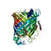
| ||||||||
|---|---|---|---|---|---|---|---|---|---|
| 1 |
| ||||||||
| Unit cell |
| ||||||||
| Details | The biological assembly is a monomer |
-Components
| #1: Protein | Mass: 31536.875 Da / Num. of mol.: 1 / Mutation: N-TERMINAL EXTENSION ARIRAP Source method: isolated from a genetically manipulated source Source: (gene. exp.) Escherichia coli (E. coli) / Description: ESCHERICHIA COLI, OUTER MEMBRANE / Production host: Escherichia coli (E. coli) / References: UniProt: P0A921, phospholipase A1 | ||
|---|---|---|---|
| #2: Sugar | ChemComp-BOG / Octyl glucoside  Type: D-saccharide / Mass: 292.369 Da / Num. of mol.: 5 / Source method: obtained synthetically / Formula: C14H28O6 / Comment: detergent*YM Type: D-saccharide / Mass: 292.369 Da / Num. of mol.: 5 / Source method: obtained synthetically / Formula: C14H28O6 / Comment: detergent*YM#3: Water | ChemComp-HOH / | Water Mass: 18.015 Da / Num. of mol.: 28 / Source method: isolated from a natural source / Formula: H2O Mass: 18.015 Da / Num. of mol.: 28 / Source method: isolated from a natural source / Formula: H2O |
-Experimental details
-Experiment
| Experiment | Method: X-RAY DIFFRACTION / Number of used crystals: 2 |
|---|
- Sample preparation
| Crystal | Density Matthews: 2.87 Å3/Da / Density % sol: 57.08 % | ||||||||||||||||||||||||||||||||||||||||||||||||
|---|---|---|---|---|---|---|---|---|---|---|---|---|---|---|---|---|---|---|---|---|---|---|---|---|---|---|---|---|---|---|---|---|---|---|---|---|---|---|---|---|---|---|---|---|---|---|---|---|---|
| Crystal grow | Temperature: 293 K / Method: vapor diffusion, hanging drop / pH: 5.9 Details: 29% MPD, 0.1 M Bis-Tris, 1 mM calciumchloride, pH 5.9, VAPOR DIFFUSION, HANGING DROP, temperature 20K | ||||||||||||||||||||||||||||||||||||||||||||||||
| Crystal grow | *PLUS pH: 6.5 / Method: vapor diffusionDetails: protein solution is mixed in a 3:2 ratio with well solution | ||||||||||||||||||||||||||||||||||||||||||||||||
| Components of the solutions | *PLUS
|
-Data collection
| Diffraction |
| |||||||||||||||
|---|---|---|---|---|---|---|---|---|---|---|---|---|---|---|---|---|
| Diffraction source |
| |||||||||||||||
| Detector |
| |||||||||||||||
| Radiation | Protocol: SINGLE WAVELENGTH / Monochromatic (M) / Laue (L): M / Scattering type: x-ray | |||||||||||||||
| Radiation wavelength |
| |||||||||||||||
| Reflection | Resolution: 2.17→37 Å / Num. all: 225025 / Num. obs: 15058 / % possible obs: 76.6 % / Redundancy: 14.9 % / Biso Wilson estimate: 44.5 Å2 / Rmerge(I) obs: 0.105 / Net I/σ(I): 15 | |||||||||||||||
| Reflection shell | Resolution: 2.17→2.25 Å / Rmerge(I) obs: 0.21 / Num. unique all: 254 / % possible all: 14.4 | |||||||||||||||
| Reflection shell | *PLUS % possible obs: 14.4 % |


- Processing
| Software |
| ||||||||||||||||||||||||||||||||||||||||||||||||||||||||||||||||||||||||||||||||
|---|---|---|---|---|---|---|---|---|---|---|---|---|---|---|---|---|---|---|---|---|---|---|---|---|---|---|---|---|---|---|---|---|---|---|---|---|---|---|---|---|---|---|---|---|---|---|---|---|---|---|---|---|---|---|---|---|---|---|---|---|---|---|---|---|---|---|---|---|---|---|---|---|---|---|---|---|---|---|---|---|---|
| Refinement | Resolution: 2.17→36.6 Å / Rfactor Rfree error: 0.007 / Data cutoff high absF: 100000 / Data cutoff low absF: 0 / Isotropic thermal model: RESTRAINED / Cross valid method: THROUGHOUT / σ(F): 0 / Stereochemistry target values: Engh & Huber Details: RESOLUTION-DEPENDENT WEIGHTING SCHEME BULK SOLVENT CORRECTION
| ||||||||||||||||||||||||||||||||||||||||||||||||||||||||||||||||||||||||||||||||
| Displacement parameters | Biso mean: 56 Å2
| ||||||||||||||||||||||||||||||||||||||||||||||||||||||||||||||||||||||||||||||||
| Refine analyze |
| ||||||||||||||||||||||||||||||||||||||||||||||||||||||||||||||||||||||||||||||||
| Refinement step | Cycle: LAST / Resolution: 2.17→36.6 Å
| ||||||||||||||||||||||||||||||||||||||||||||||||||||||||||||||||||||||||||||||||
| Refine LS restraints |
| ||||||||||||||||||||||||||||||||||||||||||||||||||||||||||||||||||||||||||||||||
| LS refinement shell | Resolution: 2.17→2.25 Å / Rfactor Rfree error: 0.078 / Total num. of bins used: 10
| ||||||||||||||||||||||||||||||||||||||||||||||||||||||||||||||||||||||||||||||||
| Xplor file |
|
