 Movie
Movie Controller

[English] 日本語
 Yorodumi
- PDB-1q7r: X-ray crystallographic analysis of a predicted amidotransferase f... -
+ Open data

- Basic information
| Entry | Database: PDB / ID: 1q7r | ||||||
|---|---|---|---|---|---|---|---|
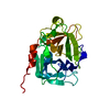
| Title | X-ray crystallographic analysis of a predicted amidotransferase from B. stearothermophilus at 1.9 A resolution | ||||||
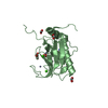
 Components Components | predicted amidotransferase | ||||||
 Keywords Keywords |  TRANSFERASE / structural genomics / yaaE / pdx2 / predicted glutamine amidotransferase / PSI / Protein Structure Initiative / Midwest Center for Structural Genomics / MCSG TRANSFERASE / structural genomics / yaaE / pdx2 / predicted glutamine amidotransferase / PSI / Protein Structure Initiative / Midwest Center for Structural Genomics / MCSG | ||||||
| Function / homology |  Function and homology information Function and homology informationpyridoxal 5'-phosphate synthase (glutamine hydrolysing) / pyridoxal 5'-phosphate synthase (glutamine hydrolysing) activity / pyridoxal phosphate biosynthetic process / glutamine catabolic process / glutaminase / glutaminase activitySimilarity search - Function | ||||||
| Biological species |   Geobacillus stearothermophilus (bacteria) Geobacillus stearothermophilus (bacteria) | ||||||
| Method | X-RAY DIFFRACTION / SYNCHROTRON / SAD / Resolution: 1.9 Å | ||||||
 Authors Authors | Miller, D.J. / Anderson, W.F. / Midwest Center for Structural Genomics (MCSG) | ||||||
 Citation Citation | Journal: To be Published Title: X-ray structure analysis of a predicted amidotransferase from B. stearothermophilus at 1.9 A resolution Authors: Miller, D.J. / Anderson, W.F. | ||||||
| History |
| ||||||
| Remark 999 | sequence No appropriate sequence database reference was available at the time of processing this file. |
- Structure visualization
| Structure viewer | Molecule: MolmilJmol/JSmol |
|---|
- Downloads & links
-Download
| PDBx/mmCIF format | 1q7r.cif.gz | 52.8 KB | Display | PDBx/mmCIF format |
|---|---|---|---|---|
| PDB format | pdb1q7r.ent.gz | 41.1 KB | Display | PDB format |
| PDBx/mmJSON format | 1q7r.json.gz | Tree view | PDBx/mmJSON format | |
| Others |  Other downloads Other downloads |
-Validation report
| Arichive directory | https://data.pdbj.org/pub/pdb/validation_reports/q7/1q7rftp://data.pdbj.org/pub/pdb/validation_reports/q7/1q7r | HTTPS FTP |
|---|
-Related structure data
| Similar structure data | |
|---|---|
| Other databases |


-Links
 PDBj
PDBj

- Assembly
| Deposited unit | 
| ||||||||
|---|---|---|---|---|---|---|---|---|---|
| 1 |
| ||||||||
| Unit cell |
|
-Components
| #1: Protein | Mass: 24439.818 Da / Num. of mol.: 1 Source method: isolated from a genetically manipulated source Source: (gene. exp.) Geobacillus stearothermophilus (bacteria)Species (production host): Escherichia coli / Production host: Escherichia coli BL21(DE3) (bacteria) / Strain (production host): BL21-DE3 / References: UniProt: P83813 |
|---|---|
| #2: Chemical | ChemComp-SO4 / Sulfate  Mass: 96.063 Da / Num. of mol.: 1 / Source method: obtained synthetically / Formula: SO4 Mass: 96.063 Da / Num. of mol.: 1 / Source method: obtained synthetically / Formula: SO4 |
| #3: Chemical | ChemComp-CL / Chloride  Mass: 35.453 Da / Num. of mol.: 1 / Source method: obtained synthetically / Formula: Cl Mass: 35.453 Da / Num. of mol.: 1 / Source method: obtained synthetically / Formula: Cl |
| #4: Chemical | ChemComp-EDO / Ethylene glycol  Mass: 62.068 Da / Num. of mol.: 1 / Source method: obtained synthetically / Formula: C2H6O2 Mass: 62.068 Da / Num. of mol.: 1 / Source method: obtained synthetically / Formula: C2H6O2 |
| #5: Water | ChemComp-HOH / Water Mass: 18.015 Da / Num. of mol.: 126 / Source method: isolated from a natural source / Formula: H2O Mass: 18.015 Da / Num. of mol.: 126 / Source method: isolated from a natural source / Formula: H2O |
-Experimental details
-Experiment
| Experiment | Method: X-RAY DIFFRACTION / Number of used crystals: 1 |
|---|
- Sample preparation
| Crystal | Density Matthews: 2.09 Å3/Da / Density % sol: 41.25 % |
|---|---|
| Crystal grow | Temperature: 277 K / Method: vapor diffusion, hanging drop / pH: 5 Details: drop: 0.05 M NaAc pH 5.0, 5 mM Tris pH 8.3, 9% PEG 4K, 0.15 M AmS, 0.25 M NaCl, 2.5 mM DTT; resevoir: 0.1 M NaAc pH 5.0, 18 % PEG 4K, 0.3 M AmS., VAPOR DIFFUSION, HANGING DROP, temperature 277K |
-Data collection
| Diffraction | Mean temperature: 170 K |
|---|---|
| Diffraction source | Source: SYNCHROTRON / Site: APS  / Beamline: 5ID-B / Wavelength: 0.979174 Å / Beamline: 5ID-B / Wavelength: 0.979174 Å |
| Detector | Type: MARRESEARCH / Detector: CCD / Date: Jun 29, 2003 |
| Radiation | Monochromator: Si 111 CHANNEL / Protocol: SAD / Monochromatic (M) / Laue (L): M / Scattering type: x-ray |
| Radiation wavelength | Wavelength: 0.979174 Å / Relative weight: 1 |
| Reflection | Resolution: 1.9→27 Å / Num. all: 16403 / Num. obs: 16323 / % possible obs: 93.4 % / Observed criterion σ(F): 0 / Observed criterion σ(I): -3 / Redundancy: 6 % / Biso Wilson estimate: 24.5 Å2 / Rmerge(I) obs: 0.083 / Net I/σ(I): 5.5 |
| Reflection shell | Resolution: 1.9→1.99 Å / Redundancy: 6.2 % / Rmerge(I) obs: 0.286 / Mean I/σ(I) obs: 2.6 / Num. unique all: 2082 / % possible all: 84.4 |
- Processing
| Software |
| ||||||||||||||||||||||||||||||||||||||||||||||||||||||||||||||||||||||||||||||||||||||||||||||||||||
|---|---|---|---|---|---|---|---|---|---|---|---|---|---|---|---|---|---|---|---|---|---|---|---|---|---|---|---|---|---|---|---|---|---|---|---|---|---|---|---|---|---|---|---|---|---|---|---|---|---|---|---|---|---|---|---|---|---|---|---|---|---|---|---|---|---|---|---|---|---|---|---|---|---|---|---|---|---|---|---|---|---|---|---|---|---|---|---|---|---|---|---|---|---|---|---|---|---|---|---|---|---|
| Refinement | Method to determine structure: SAD / Resolution: 1.9→19.73 Å / Cor.coef. Fo:Fc: 0.951 / Cor.coef. Fo:Fc free: 0.939 / SU B: 3.548 / SU ML: 0.104 / Cross valid method: THROUGHOUT / σ(F): 0 / ESU R: 0.167 / ESU R Free: 0.151 / Stereochemistry target values: MAXIMUM LIKELIHOOD / Details: HYDROGENS HAVE BEEN ADDED IN THE RIDING POSITIONS
| ||||||||||||||||||||||||||||||||||||||||||||||||||||||||||||||||||||||||||||||||||||||||||||||||||||
| Solvent computation | Ion probe radii: 0.8 Å / Shrinkage radii: 0.8 Å / VDW probe radii: 1.4 Å / Solvent model: BABINET MODEL WITH MASK | ||||||||||||||||||||||||||||||||||||||||||||||||||||||||||||||||||||||||||||||||||||||||||||||||||||
| Displacement parameters | Biso mean: 33.002 Å2
| ||||||||||||||||||||||||||||||||||||||||||||||||||||||||||||||||||||||||||||||||||||||||||||||||||||
| Refinement step | Cycle: LAST / Resolution: 1.9→19.73 Å
| ||||||||||||||||||||||||||||||||||||||||||||||||||||||||||||||||||||||||||||||||||||||||||||||||||||
| Refine LS restraints |
| ||||||||||||||||||||||||||||||||||||||||||||||||||||||||||||||||||||||||||||||||||||||||||||||||||||
| LS refinement shell | Resolution: 1.9→1.949 Å / Total num. of bins used: 20
|
