 Movie
Movie Controller


+ Open data

- Basic information
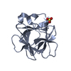
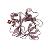
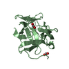
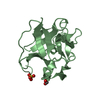
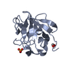
| Entry | Database: PDB / ID: 1q03 | ||||||
|---|---|---|---|---|---|---|---|
| Title | Crystal structure of FGF-1, S50G/V51G mutant | ||||||
 Components Components | Heparin-binding growth factor 1 | ||||||
 Keywords Keywords | hormone/growth factor / beta-trefoil / hormone-growth factor COMPLEX | ||||||
| Function / homology |  Function and homology information Function and homology informationmesonephric epithelium development / branch elongation involved in ureteric bud branching / regulation of endothelial tube morphogenesis / FGFR3b ligand binding and activation / regulation of endothelial cell chemotaxis to fibroblast growth factor / Signaling by activated point mutants of FGFR3 / FGFR3c ligand binding and activation / Phospholipase C-mediated cascade; FGFR3 / FGFR2b ligand binding and activation / positive regulation of cholesterol biosynthetic process ...mesonephric epithelium development / branch elongation involved in ureteric bud branching / regulation of endothelial tube morphogenesis / FGFR3b ligand binding and activation / regulation of endothelial cell chemotaxis to fibroblast growth factor / Signaling by activated point mutants of FGFR3 / FGFR3c ligand binding and activation / Phospholipase C-mediated cascade; FGFR3 / FGFR2b ligand binding and activation / positive regulation of cholesterol biosynthetic process /  fibroblast growth factor receptor binding / FGFR2c ligand binding and activation / Activated point mutants of FGFR2 / Phospholipase C-mediated cascade; FGFR2 / FGFR4 ligand binding and activation / FGFR1b ligand binding and activation / Phospholipase C-mediated cascade; FGFR4 / Signaling by activated point mutants of FGFR1 / FGFR1c ligand binding and activation / organ induction / Downstream signaling of activated FGFR1 / Phospholipase C-mediated cascade: FGFR1 / positive regulation of hepatocyte proliferation / S100 protein binding / positive regulation of intracellular signal transduction / Signaling by FGFR2 IIIa TM / PI-3K cascade:FGFR3 / positive regulation of sprouting angiogenesis / PI-3K cascade:FGFR2 / PI-3K cascade:FGFR4 / PI-3K cascade:FGFR1 / positive regulation of cell division / PI3K Cascade / anatomical structure morphogenesis / fibroblast growth factor receptor signaling pathway / SHC-mediated cascade:FGFR3 / SHC-mediated cascade:FGFR2 / SHC-mediated cascade:FGFR4 / SHC-mediated cascade:FGFR1 / FRS-mediated FGFR3 signaling / FRS-mediated FGFR2 signaling / FRS-mediated FGFR4 signaling / Signaling by FGFR3 in disease / FRS-mediated FGFR1 signaling / Hsp70 protein binding / Signaling by FGFR2 in disease / Signaling by FGFR1 in disease / activation of protein kinase B activity / extracellular matrix / positive regulation of endothelial cell migration / epithelial cell proliferation / positive regulation of epithelial cell proliferation / Negative regulation of FGFR3 signaling / Negative regulation of FGFR2 signaling / Negative regulation of FGFR4 signaling / Negative regulation of FGFR1 signaling / animal organ morphogenesis / lung development / growth factor activity / positive regulation of MAP kinase activity / wound healing / positive regulation of angiogenesis / Constitutive Signaling by Aberrant PI3K in Cancer / integrin binding / PIP3 activates AKT signaling / cellular response to heat / heparin binding / cell cortex / PI5P, PP2A and IER3 Regulate PI3K/AKT Signaling / RAF/MAP kinase cascade / angiogenesis / cell differentiation / positive regulation of ERK1 and ERK2 cascade / positive regulation of cell migration / positive regulation of cell population proliferation / positive regulation of gene expression / signal transduction / positive regulation of transcription by RNA polymerase II / extracellular space / extracellular region / nucleoplasm / nucleus / cytosol / cytoplasm fibroblast growth factor receptor binding / FGFR2c ligand binding and activation / Activated point mutants of FGFR2 / Phospholipase C-mediated cascade; FGFR2 / FGFR4 ligand binding and activation / FGFR1b ligand binding and activation / Phospholipase C-mediated cascade; FGFR4 / Signaling by activated point mutants of FGFR1 / FGFR1c ligand binding and activation / organ induction / Downstream signaling of activated FGFR1 / Phospholipase C-mediated cascade: FGFR1 / positive regulation of hepatocyte proliferation / S100 protein binding / positive regulation of intracellular signal transduction / Signaling by FGFR2 IIIa TM / PI-3K cascade:FGFR3 / positive regulation of sprouting angiogenesis / PI-3K cascade:FGFR2 / PI-3K cascade:FGFR4 / PI-3K cascade:FGFR1 / positive regulation of cell division / PI3K Cascade / anatomical structure morphogenesis / fibroblast growth factor receptor signaling pathway / SHC-mediated cascade:FGFR3 / SHC-mediated cascade:FGFR2 / SHC-mediated cascade:FGFR4 / SHC-mediated cascade:FGFR1 / FRS-mediated FGFR3 signaling / FRS-mediated FGFR2 signaling / FRS-mediated FGFR4 signaling / Signaling by FGFR3 in disease / FRS-mediated FGFR1 signaling / Hsp70 protein binding / Signaling by FGFR2 in disease / Signaling by FGFR1 in disease / activation of protein kinase B activity / extracellular matrix / positive regulation of endothelial cell migration / epithelial cell proliferation / positive regulation of epithelial cell proliferation / Negative regulation of FGFR3 signaling / Negative regulation of FGFR2 signaling / Negative regulation of FGFR4 signaling / Negative regulation of FGFR1 signaling / animal organ morphogenesis / lung development / growth factor activity / positive regulation of MAP kinase activity / wound healing / positive regulation of angiogenesis / Constitutive Signaling by Aberrant PI3K in Cancer / integrin binding / PIP3 activates AKT signaling / cellular response to heat / heparin binding / cell cortex / PI5P, PP2A and IER3 Regulate PI3K/AKT Signaling / RAF/MAP kinase cascade / angiogenesis / cell differentiation / positive regulation of ERK1 and ERK2 cascade / positive regulation of cell migration / positive regulation of cell population proliferation / positive regulation of gene expression / signal transduction / positive regulation of transcription by RNA polymerase II / extracellular space / extracellular region / nucleoplasm / nucleus / cytosol / cytoplasmSimilarity search - Function | ||||||
| Biological species |  Homo sapiens (human) Homo sapiens (human) | ||||||
| Method | X-RAY DIFFRACTION / MOLECULAR REPLACEMENT / Resolution: 2.05 Å | ||||||
 Authors Authors | Kim, J. / Blaber, M. | ||||||
 Citation Citation | Journal: Protein Sci. / Year: 2005 Title: Sequence swapping does not result in conformation swapping for the beta4/beta5 and beta8/beta9 beta-hairpin turns in human acidic fibroblast growth factor Authors: Kim, J. / Lee, J. / Brych, S.R. / Logan, T.M. / Blaber, M. | ||||||
| History |
|
- Structure visualization
| Structure viewer | Molecule: MolmilJmol/JSmol |
|---|
- Downloads & links
-Download
| PDBx/mmCIF format | 1q03.cif.gz | 69.1 KB | Display | PDBx/mmCIF format |
|---|---|---|---|---|
| PDB format | pdb1q03.ent.gz | 51.9 KB | Display | PDB format |
| PDBx/mmJSON format | 1q03.json.gz | Tree view | PDBx/mmJSON format | |
| Others |  Other downloads Other downloads |
-Validation report
| Arichive directory | https://data.pdbj.org/pub/pdb/validation_reports/q0/1q03ftp://data.pdbj.org/pub/pdb/validation_reports/q0/1q03 | HTTPS FTP |
|---|
-Related structure data
| Related structure data |  1pzzC  1q04C 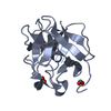 1jqzS S: Starting model for refinement C: citing same article ( |
|---|---|
| Similar structure data |


-Links
 PDBj
PDBj


- Assembly
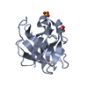
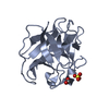
| Deposited unit | 
| ||||||||||
|---|---|---|---|---|---|---|---|---|---|---|---|
| 1 | 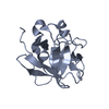
| ||||||||||
| 2 | 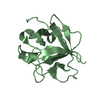
| ||||||||||
| Unit cell |
|
-Components
| #1: Protein | Mass: 16614.643 Da / Num. of mol.: 2 / Mutation: S50G, V51G Source method: isolated from a genetically manipulated source Source: (gene. exp.) Homo sapiens (human) / Production host:  Escherichia coli (E. coli) / References: UniProt: P05230 Escherichia coli (E. coli) / References: UniProt: P05230#2: Water | ChemComp-HOH / | Water Mass: 18.015 Da / Num. of mol.: 129 / Source method: isolated from a natural source / Formula: H2O Mass: 18.015 Da / Num. of mol.: 129 / Source method: isolated from a natural source / Formula: H2O |
|---|
-Experimental details
-Experiment
| Experiment | Method: X-RAY DIFFRACTION / Number of used crystals: 1 |
|---|
- Sample preparation
| Crystal | Density Matthews: 2.94 Å3/Da / Density % sol: 57.8 % |
|---|---|
| Crystal grow | Temperature: 298.15 K / Method: vapor diffusion, hanging drop / pH: 7.5 Details: Formate, pH 7.5, VAPOR DIFFUSION, HANGING DROP, temperature 298.15K |
-Data collection
| Diffraction source | Source: ROTATING ANODE / Type: RIGAKU / Wavelength: 1.5418 Å |
|---|---|
| Detector | Type: MARRESEARCH / Detector: CCD / Date: Jan 1, 2003 / Details: Osmic Purple confocal mirror |
| Radiation | Protocol: SINGLE WAVELENGTH / Monochromatic (M) / Laue (L): M / Scattering type: x-ray |
| Radiation wavelength | Wavelength: 1.5418 Å / Relative weight: 1 |
| Reflection | Resolution: 2.05→19.44 Å / Num. all: 25055 / Num. obs: 20665 / % possible obs: 82.5 % / Observed criterion σ(F): 3 / Observed criterion σ(I): 1.5 / Rmerge(I) obs: 0.079 |
| Reflection shell | Resolution: 2.05→2.1 Å / Rmerge(I) obs: 0.398 / Mean I/σ(I) obs: 4.7 / % possible all: 100 |
- Processing
| Software |
| |||||||||||||||||||||||||
|---|---|---|---|---|---|---|---|---|---|---|---|---|---|---|---|---|---|---|---|---|---|---|---|---|---|---|
| Refinement | Method to determine structure: MOLECULAR REPLACEMENT Starting model: 1JQZ Resolution: 2.05→19.44 Å / Cross valid method: THROUGHOUT / σ(F): 3
| |||||||||||||||||||||||||
| Refinement step | Cycle: LAST / Resolution: 2.05→19.44 Å
| |||||||||||||||||||||||||
| Refine LS restraints |
| |||||||||||||||||||||||||
| LS refinement shell | Resolution: 2.05→2.1 Å / Rfactor Rfree error: 0.021
|
