 Movie
Movie Controller

[English] 日本語
 Yorodumi
- PDB-1py1: Complex of GGA1-VHS domain and beta-secretase C-terminal phosphop... -
+ Open data

- Basic information
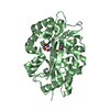
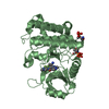
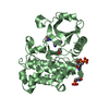
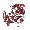
| Entry | Database: PDB / ID: 1py1 | ||||||
|---|---|---|---|---|---|---|---|
| Title | Complex of GGA1-VHS domain and beta-secretase C-terminal phosphopeptide | ||||||
 Components Components |
| ||||||
 Keywords Keywords |  PROTEIN TRANSPORT / VHS DOMAIN OF GGA1 / BETA-SECRETASE / PROTEIN-PEPTIDE COMPLEX / SUPER HELIX PROTEIN TRANSPORT / VHS DOMAIN OF GGA1 / BETA-SECRETASE / PROTEIN-PEPTIDE COMPLEX / SUPER HELIX | ||||||
| Function / homology |  Function and homology information Function and homology informationprotein localization to ciliary membrane / Golgi to plasma membrane protein transport / Golgi to plasma membrane transport / protein localization to cell surface / retrograde transport, endosome to Golgi / TBC/RABGAPs / memapsin 2 / Golgi-associated vesicle lumen / signaling receptor ligand precursor processing / beta-aspartyl-peptidase activity ...protein localization to ciliary membrane / Golgi to plasma membrane protein transport / Golgi to plasma membrane transport / protein localization to cell surface / retrograde transport, endosome to Golgi / TBC/RABGAPs / memapsin 2 / Golgi-associated vesicle lumen / signaling receptor ligand precursor processing / beta-aspartyl-peptidase activity / amyloid precursor protein catabolic process / amyloid-beta formation / membrane protein ectodomain proteolysis / cellular response to manganese ion / prepulse inhibition / amyloid-beta metabolic process / detection of mechanical stimulus involved in sensory perception of pain / cellular response to copper ion / hippocampal mossy fiber to CA3 synapse / presynaptic modulation of chemical synaptic transmission / multivesicular body / phosphatidylinositol binding / ubiquitin binding / response to lead ion / intracellular protein transport / protein catabolic process / trans-Golgi network / protein localization / protein processing / recycling endosome / small GTPase binding / cellular response to amyloid-beta / positive regulation of protein catabolic process / positive regulation of neuron apoptotic process / synaptic vesicle / late endosome / amyloid-beta binding / peptidase activity / early endosome membrane / endopeptidase activity / amyloid fibril formation / lysosome / aspartic-type endopeptidase activity / early endosome / endosome membrane / endosome / membrane raft / Amyloid fiber formation / axon / endoplasmic reticulum lumen / intracellular membrane-bounded organelle / neuronal cell body / dendrite / Golgi apparatus / enzyme binding / cell surface / protein-containing complex / proteolysis / nucleoplasm / membrane / plasma membrane / cytosolSimilarity search - Function | ||||||
| Biological species |  Homo sapiens (human) Homo sapiens (human) | ||||||
| Method | X-RAY DIFFRACTION / MOLECULAR REPLACEMENT / Resolution: 2.6 Å | ||||||
 Authors Authors | Zhu, G. / Zhang, X.C. | ||||||
 Citation Citation | Journal: Biochemistry / Year: 2003 Title: Biochemical and structural characterization of the interaction of memapsin 2 (beta-secretase) cytosolic domain with the VHS domain of GGA proteins. Authors: He, X. / Zhu, G. / Koelsch, G. / Rodgers, K. / Zhang, X.C. / Tang, J. | ||||||
| History |
|
- Structure visualization
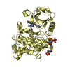
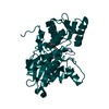
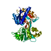
| Structure viewer | Molecule: MolmilJmol/JSmol |
|---|
- Downloads & links
-Download
| PDBx/mmCIF format | 1py1.cif.gz | 126.7 KB | Display | PDBx/mmCIF format |
|---|---|---|---|---|
| PDB format | pdb1py1.ent.gz | 101.1 KB | Display | PDB format |
| PDBx/mmJSON format | 1py1.json.gz | Tree view | PDBx/mmJSON format | |
| Others |  Other downloads Other downloads |
-Validation report
| Arichive directory | https://data.pdbj.org/pub/pdb/validation_reports/py/1py1ftp://data.pdbj.org/pub/pdb/validation_reports/py/1py1 | HTTPS FTP |
|---|
-Related structure data
| Related structure data | 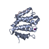 1jwgS S: Starting model for refinement |
|---|---|
| Similar structure data |


-Links
 PDBj
PDBj


- Assembly
| Deposited unit | 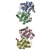
| ||||||||
|---|---|---|---|---|---|---|---|---|---|
| 1 | 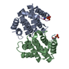
| ||||||||
| 2 | 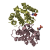
| ||||||||
| Unit cell |
|
-Components
| #1: Protein | Mass: 17958.586 Da / Num. of mol.: 4 / Fragment: VHS Domain (Residues 2-157) Source method: isolated from a genetically manipulated source Source: (gene. exp.) Homo sapiens (human) / Gene: GGA1 / Plasmid: pGEX2T / Species (production host): Escherichia coli / Production host:  Escherichia coli BL21(DE3) (bacteria) / Strain (production host): BL21(DE3) / References: UniProt: Q9UJY5 Escherichia coli BL21(DE3) (bacteria) / Strain (production host): BL21(DE3) / References: UniProt: Q9UJY5#2: Protein/peptide | / Beta-site APP cleaving enzyme / Beta-site amyloid precursor protein cleaving enzyme / Aspartyl ...Beta-site APP cleaving enzyme / Beta-site amyloid precursor protein cleaving enzyme / Aspartyl protease 2 / Asp 2 / ASP2 / Membrane-associated aspartic protease 2 / Memapsin-2Mass: 954.979 Da / Num. of mol.: 4 / Fragment: C-TERMINUS (RESIDUES 494-501) / Source method: obtained synthetically / Details: YES References: UniProt: P56817, Hydrolases; Acting on peptide bonds (peptidases); Aspartic endopeptidases#3: Water | ChemComp-HOH / | Water Mass: 18.015 Da / Num. of mol.: 37 / Source method: isolated from a natural source / Formula: H2O Mass: 18.015 Da / Num. of mol.: 37 / Source method: isolated from a natural source / Formula: H2O |
|---|
-Experimental details
-Experiment
| Experiment | Method: X-RAY DIFFRACTION / Number of used crystals: 1 |
|---|
- Sample preparation
| Crystal | Density Matthews: 2.39 Å3/Da / Density % sol: 48.46 % | ||||||||||||||||||||||||||||||
|---|---|---|---|---|---|---|---|---|---|---|---|---|---|---|---|---|---|---|---|---|---|---|---|---|---|---|---|---|---|---|---|
| Crystal grow | Temperature: 293 K / Method: vapor diffusion, hanging drop / pH: 6.4 Details: PEG 3350, AMMONIUM SULFATE, CACODYLATE, pH 6.4, VAPOR DIFFUSION, HANGING DROP, temperature 293K | ||||||||||||||||||||||||||||||
| Crystal grow | *PLUS Temperature: 20 ℃ / Method: vapor diffusion, hanging drop | ||||||||||||||||||||||||||||||
| Components of the solutions | *PLUS
|
-Data collection
| Diffraction | Mean temperature: 100 K |
|---|---|
| Diffraction source | Source: ROTATING ANODE / Type: RIGAKU RU300 / Wavelength: 1.5418 Å |
| Detector | Type: MARRESEARCH / Detector: IMAGE PLATE / Date: Mar 17, 2003 |
| Radiation | Monochromator: OSMIC OPTICS / Protocol: SINGLE WAVELENGTH / Monochromatic (M) / Laue (L): M / Scattering type: x-ray |
| Radiation wavelength | Wavelength: 1.5418 Å / Relative weight: 1 |
| Reflection | Resolution: 2.6→40 Å / Num. all: 22869 / Num. obs: 22025 / % possible obs: 95.8 % / Observed criterion σ(F): 0 / Observed criterion σ(I): 0 / Redundancy: 6.9 % / Biso Wilson estimate: 46.9 Å2 / Rmerge(I) obs: 0.074 / Net I/σ(I): 25.8 |
| Reflection shell | Resolution: 2.6→2.69 Å / Rmerge(I) obs: 0.373 / Mean I/σ(I) obs: 4 / Num. unique all: 2229 / % possible all: 98.9 |
| Reflection | *PLUS Lowest resolution: 40 Å / Num. obs: 22869 / % possible obs: 99.5 % |
| Reflection shell | *PLUS % possible obs: 98.9 % / Num. unique obs: 2229 / Rmerge(I) obs: 0.477 / Mean I/σ(I) obs: 3.6 |
- Processing
| Software |
| ||||||||||||||||||||||||||||||||||||
|---|---|---|---|---|---|---|---|---|---|---|---|---|---|---|---|---|---|---|---|---|---|---|---|---|---|---|---|---|---|---|---|---|---|---|---|---|---|
| Refinement | Method to determine structure: MOLECULAR REPLACEMENT Starting model: PDB ENTRY 1JWG Resolution: 2.6→39.45 Å / Rfactor Rfree error: 0.01 / Isotropic thermal model: RESTRAINED / Cross valid method: THROUGHOUT / σ(F): 0 / Stereochemistry target values: Engh & Huber
| ||||||||||||||||||||||||||||||||||||
| Solvent computation | Solvent model: FLAT MODEL / Bsol: 25.6879 Å2 / ksol: 0.307058 e/Å3 | ||||||||||||||||||||||||||||||||||||
| Displacement parameters | Biso mean: 52.5 Å2
| ||||||||||||||||||||||||||||||||||||
| Refine analyze | Luzzati coordinate error free: 0.51 Å / Luzzati sigma a free: 0.53 Å | ||||||||||||||||||||||||||||||||||||
| Refinement step | Cycle: LAST / Resolution: 2.6→39.45 Å
| ||||||||||||||||||||||||||||||||||||
| Refine LS restraints |
| ||||||||||||||||||||||||||||||||||||
| LS refinement shell | Resolution: 2.6→2.69 Å / Rfactor Rfree error: 0.062 / Total num. of bins used: 10
| ||||||||||||||||||||||||||||||||||||
| Xplor file |
| ||||||||||||||||||||||||||||||||||||
| Refinement | *PLUS Lowest resolution: 40 Å | ||||||||||||||||||||||||||||||||||||
| Solvent computation | *PLUS | ||||||||||||||||||||||||||||||||||||
| Displacement parameters | *PLUS | ||||||||||||||||||||||||||||||||||||
| Refine LS restraints | *PLUS
|
