 Movie
Movie Controller


+ Open data

- Basic information
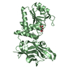
| Entry | Database: PDB / ID: 1puc | ||||||
|---|---|---|---|---|---|---|---|
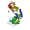
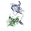
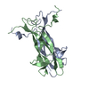
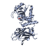
| Title | P13SUC1 IN A STRAND-EXCHANGED DIMER | ||||||
 Components Components | P13SUC1 | ||||||
 Keywords Keywords |  BINDING PROTEIN / CELL CYCLE / DOMAIN SWAPPING / STRAND-EXCHANGED DIMER BINDING PROTEIN / CELL CYCLE / DOMAIN SWAPPING / STRAND-EXCHANGED DIMER | ||||||
| Function / homology |  Function and homology informationsignaling / cyclin-dependent protein serine/threonine kinase activator activity / SCF ubiquitin ligase complex / cyclin-dependent protein kinase holoenzyme complex / regulation of mitotic cell cycle / ubiquitin binding / histone binding / cell cycle / cell division / protein-containing complex binding ...signaling / cyclin-dependent protein serine/threonine kinase activator activity / SCF ubiquitin ligase complex / cyclin-dependent protein kinase holoenzyme complex / regulation of mitotic cell cycle / ubiquitin binding / histone binding / cell cycle / cell division / protein-containing complex binding / protein kinase binding / zinc ion binding / nucleus / cytosol Function and homology informationsignaling / cyclin-dependent protein serine/threonine kinase activator activity / SCF ubiquitin ligase complex / cyclin-dependent protein kinase holoenzyme complex / regulation of mitotic cell cycle / ubiquitin binding / histone binding / cell cycle / cell division / protein-containing complex binding ...signaling / cyclin-dependent protein serine/threonine kinase activator activity / SCF ubiquitin ligase complex / cyclin-dependent protein kinase holoenzyme complex / regulation of mitotic cell cycle / ubiquitin binding / histone binding / cell cycle / cell division / protein-containing complex binding / protein kinase binding / zinc ion binding / nucleus / cytosolSimilarity search - Function | ||||||
| Biological species |  Schizosaccharomyces pombe (fission yeast) Schizosaccharomyces pombe (fission yeast) | ||||||
| Method | X-RAY DIFFRACTION / Resolution: 1.95 Å | ||||||
 Authors Authors | Khazanovich, N. / Bateman, K.S. / Chernaia, M. / Michalak, M. / James, M.N.G. | ||||||
 Citation Citation | Journal: Structure / Year: 1996 Title: Crystal structure of the yeast cell-cycle control protein, p13suc1, in a strand-exchanged dimer. Authors: Khazanovich, N. / Bateman, K. / Chernaia, M. / Michalak, M. / James, M. | ||||||
| History |
| ||||||
| Remark 700 | SHEET EACH MOLECULE OF P13SUC1 IN THIS CRYSTAL PARTICIPATES IN A STRAND-EXCHANGED DIMER WITH A ...SHEET EACH MOLECULE OF P13SUC1 IN THIS CRYSTAL PARTICIPATES IN A STRAND-EXCHANGED DIMER WITH A SYMMETRY-RELATED MOLECULE. A SINGLE BETA-STRAND, ILE 94 - ASP 102, IS EXCHANGED BETWEEN THE CENTRAL BETA-SHEETS OF EACH MOLECULE. |
- Structure visualization
| Structure viewer | Molecule: MolmilJmol/JSmol |
|---|
- Downloads & links
-Download
| PDBx/mmCIF format | 1puc.cif.gz | 35.9 KB | Display | PDBx/mmCIF format |
|---|---|---|---|---|
| PDB format | pdb1puc.ent.gz | 23.7 KB | Display | PDB format |
| PDBx/mmJSON format | 1puc.json.gz | Tree view | PDBx/mmJSON format | |
| Others |  Other downloads Other downloads |
-Validation report
| Arichive directory | https://data.pdbj.org/pub/pdb/validation_reports/pu/1pucftp://data.pdbj.org/pub/pdb/validation_reports/pu/1puc | HTTPS FTP |
|---|
-Related structure data
| Similar structure data |
|---|


-Links
 PDBj
PDBj- Assembly
| Deposited unit | 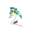
| ||||||||
|---|---|---|---|---|---|---|---|---|---|
| 1 | 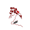
| ||||||||
| Unit cell |
|
-Components
| #1: Protein | Mass: 12572.309 Da / Num. of mol.: 1 Source method: isolated from a genetically manipulated source Source: (gene. exp.) Schizosaccharomyces pombe (fission yeast)Cell line: BL21 / Plasmid: BL21 / Species (production host): Escherichia coli / Production host:  Escherichia coli BL21(DE3) (bacteria) / Strain (production host): BL21 (DE3) / References: UniProt: P08463 Escherichia coli BL21(DE3) (bacteria) / Strain (production host): BL21 (DE3) / References: UniProt: P08463 |
|---|---|
| #2: Chemical | ChemComp-CPS / CHAPS detergent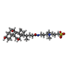  Mass: 614.877 Da / Num. of mol.: 1 / Source method: obtained synthetically / Formula: C32H58N2O7S / Comment: detergent*YM Mass: 614.877 Da / Num. of mol.: 1 / Source method: obtained synthetically / Formula: C32H58N2O7S / Comment: detergent*YM |
| #3: Water | ChemComp-HOH / Water Mass: 18.015 Da / Num. of mol.: 72 / Source method: isolated from a natural source / Formula: H2O Mass: 18.015 Da / Num. of mol.: 72 / Source method: isolated from a natural source / Formula: H2O |
-Experimental details
-Experiment
| Experiment | Method: X-RAY DIFFRACTION / Number of used crystals: 1 |
|---|
- Sample preparation
| Crystal | Density Matthews: 2.66 Å3/Da / Density % sol: 53.71 % | ||||||||||||||||||||||||||||||||||||||||||||||||||||||
|---|---|---|---|---|---|---|---|---|---|---|---|---|---|---|---|---|---|---|---|---|---|---|---|---|---|---|---|---|---|---|---|---|---|---|---|---|---|---|---|---|---|---|---|---|---|---|---|---|---|---|---|---|---|---|---|
| Crystal grow | *PLUS pH: 7.8 / Method: vapor diffusion, sitting drop | ||||||||||||||||||||||||||||||||||||||||||||||||||||||
| Components of the solutions | *PLUS
|
-Data collection
| Diffraction source | Wavelength: 1.5418 Å |
|---|---|
| Detector | Type: XUONG-HAMLIN MULTIWIRE / Detector: AREA DETECTOR / Date: Sep 28, 1994 |
| Radiation | Monochromatic (M) / Laue (L): M / Scattering type: x-ray |
| Radiation wavelength | Wavelength: 1.5418 Å / Relative weight: 1 |
| Reflection | Resolution: 1.95→55.9 Å / Num. obs: 9859 / % possible obs: 97.5 % / Redundancy: 5.4 % / Rmerge(I) obs: 0.0488 |
| Reflection | *PLUS Lowest resolution: 50 Å / Num. measured all: 53600 / Rmerge(I) obs: 0.0488 |
| Reflection shell | *PLUS Highest resolution: 1.95 Å / Lowest resolution: 1.98 Å / % possible obs: 81.4 % |
- Processing
| Software |
| ||||||||||||||||||||||||||||||
|---|---|---|---|---|---|---|---|---|---|---|---|---|---|---|---|---|---|---|---|---|---|---|---|---|---|---|---|---|---|---|---|
| Refinement | Resolution: 1.95→6 Å / σ(F): 0
| ||||||||||||||||||||||||||||||
| Displacement parameters | Biso mean: 24 Å2 | ||||||||||||||||||||||||||||||
| Refine analyze | Luzzati coordinate error obs: 0.2 Å | ||||||||||||||||||||||||||||||
| Refinement step | Cycle: LAST / Resolution: 1.95→6 Å
| ||||||||||||||||||||||||||||||
| Refine LS restraints |
| ||||||||||||||||||||||||||||||
| Software | *PLUS Name: TNT / Classification: refinement | ||||||||||||||||||||||||||||||
| Refine LS restraints | *PLUS
|
