 Movie
Movie Controller

[English] 日本語
 Yorodumi
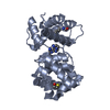
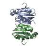
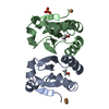
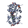
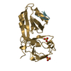
- PDB-1pu6: Crystal structure of H.pylori 3-methyladenine DNA glycosylase (MagIII) -
+ Open data

- Basic information
| Entry | Database: PDB / ID: 1pu6 | ||||||
|---|---|---|---|---|---|---|---|
| Title | Crystal structure of H.pylori 3-methyladenine DNA glycosylase (MagIII) | ||||||
 Components Components | 3-METHYLADENINE DNA GLYCOSYLASE | ||||||
 Keywords Keywords |  HYDROLASE / Helix-hairpin-helix / 3-Methyladenine / BASE EXCISION REPAIR / GLYCOSYLASE HYDROLASE / Helix-hairpin-helix / 3-Methyladenine / BASE EXCISION REPAIR / GLYCOSYLASE | ||||||
| Function / homology |  Function and homology informationbase-excision repair / 4 iron, 4 sulfur cluster binding / endonuclease activity / metal ion binding Function and homology informationbase-excision repair / 4 iron, 4 sulfur cluster binding / endonuclease activity / metal ion bindingSimilarity search - Function | ||||||
| Biological species |   Helicobacter pylori (bacteria) Helicobacter pylori (bacteria) | ||||||
| Method | X-RAY DIFFRACTION / SYNCHROTRON / MAD / Resolution: 1.64 Å | ||||||
 Authors Authors | Eichman, B.F. / O'Rourke, E.J. / Radicella, J.P. / Ellenberger, T. | ||||||
 Citation Citation | Journal: Embo J. / Year: 2003 Title: Crystal structures of 3-methyladenine DNA glycosylase MagIII and the recognition of alkylated bases Authors: Eichman, B.F. / O'Rourke, E.J. / Radicella, J.P. / Ellenberger, T. #1: Journal: J.Biol.Chem. / Year: 2000Title: A NOVEL 3-METHYLADENINE DNA GLYCOSYLASE FROM HELICOBACTER PYLORI DEFINES A NEW CLASS WITHIN THE ENDONUCLEASE III FAMILY OF BASE EXCISION REPAIR GLYCOSYLASES Authors: O'ROURKE, E.J. / CHEVALIER, C. / BOITEUX, S. / LABIGNE, A. / IELPI, L. / RADICELLA, J.P. | ||||||
| History |
| ||||||
| Remark 999 | SEQUENCE THE DIFFERENCES BETWEEN THE SEQUENCES IN THE PDB ENTRY 1PU6 AND IN THE DATABASE REFERENCE ...SEQUENCE THE DIFFERENCES BETWEEN THE SEQUENCES IN THE PDB ENTRY 1PU6 AND IN THE DATABASE REFERENCE ARE DUE TO THE PARTICULAR H.PYLORI STRAIN. DATABASE REFERENCE REFERS TO H.PYLORI STRAIN J99; MAGIII WAS CLONED FROM H.PYLORI STRAIN 13/5 (SEE REFERENCE 1) |
- Structure visualization
| Structure viewer | Molecule: MolmilJmol/JSmol |
|---|
- Downloads & links
-Download
| PDBx/mmCIF format | 1pu6.cif.gz | 187.2 KB | Display | PDBx/mmCIF format |
|---|---|---|---|---|
| PDB format | pdb1pu6.ent.gz | 157.7 KB | Display | PDB format |
| PDBx/mmJSON format | 1pu6.json.gz | Tree view | PDBx/mmJSON format | |
| Others |  Other downloads Other downloads |
-Validation report
| Arichive directory | https://data.pdbj.org/pub/pdb/validation_reports/pu/1pu6ftp://data.pdbj.org/pub/pdb/validation_reports/pu/1pu6 | HTTPS FTP |
|---|
-Related structure data


-Links
 PDBj
PDBj


- Assembly
| Deposited unit | 
| ||||||||
|---|---|---|---|---|---|---|---|---|---|
| 1 | 
| ||||||||
| 2 | 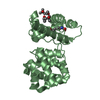
| ||||||||
| Unit cell |
|
-Components
| #1: Protein | Mass: 25218.064 Da / Num. of mol.: 2 Source method: isolated from a genetically manipulated source Source: (gene. exp.) Helicobacter pylori (bacteria) / Strain: 13-5 / Production host: Escherichia coli (E. coli) / References: UniProt: O25323#2: Chemical | ChemComp-CL / | Chloride  Mass: 35.453 Da / Num. of mol.: 1 / Source method: obtained synthetically / Formula: Cl Mass: 35.453 Da / Num. of mol.: 1 / Source method: obtained synthetically / Formula: Cl#3: Chemical | ChemComp-BME / | 2-Mercaptoethanol  Mass: 78.133 Da / Num. of mol.: 1 / Source method: obtained synthetically / Formula: C2H6OS Mass: 78.133 Da / Num. of mol.: 1 / Source method: obtained synthetically / Formula: C2H6OS#4: Chemical | 2-Methyl-2,4-pentanediol  Mass: 118.174 Da / Num. of mol.: 2 / Source method: obtained synthetically / Formula: C6H14O2 / Comment: precipitant*YM Mass: 118.174 Da / Num. of mol.: 2 / Source method: obtained synthetically / Formula: C6H14O2 / Comment: precipitant*YM#5: Water | ChemComp-HOH / | Water Mass: 18.015 Da / Num. of mol.: 234 / Source method: isolated from a natural source / Formula: H2O Mass: 18.015 Da / Num. of mol.: 234 / Source method: isolated from a natural source / Formula: H2O |
|---|
-Experimental details
-Experiment
| Experiment | Method: X-RAY DIFFRACTION / Number of used crystals: 2 |
|---|
- Sample preparation
| Crystal | Density Matthews: 2.52 Å3/Da / Density % sol: 51.24 % | |||||||||||||||
|---|---|---|---|---|---|---|---|---|---|---|---|---|---|---|---|---|
| Crystal grow | Temperature: 277 K / Method: vapor diffusion, hanging drop / pH: 7 Details: PEG 4000, HEPES, MPD, pH 7.0, VAPOR DIFFUSION, HANGING DROP, temperature 277K | |||||||||||||||
| Crystal grow | *PLUS Temperature: 4 ℃ / Method: vapor diffusion | |||||||||||||||
| Components of the solutions | *PLUS
|
-Data collection
| Diffraction |
| |||||||||||||||
|---|---|---|---|---|---|---|---|---|---|---|---|---|---|---|---|---|
| Diffraction source |
| |||||||||||||||
| Detector |
| |||||||||||||||
| Radiation |
| |||||||||||||||
| Radiation wavelength |
| |||||||||||||||
| Reflection | Resolution: 1.64→50 Å / Num. all: 60643 / Num. obs: 60643 / % possible obs: 99.9 % / Observed criterion σ(F): 0 / Observed criterion σ(I): 0 / Redundancy: 8.1 % / Biso Wilson estimate: 19.5 Å2 / Rsym value: 0.099 / Net I/σ(I): 21.8 | |||||||||||||||
| Reflection shell | Resolution: 1.64→1.71 Å / Redundancy: 5.7 % / Mean I/σ(I) obs: 4.3 / Rsym value: 0.337 / % possible all: 99.7 | |||||||||||||||
| Reflection | *PLUS Lowest resolution: 25 Å / Rmerge(I) obs: 0.099 | |||||||||||||||
| Reflection shell | *PLUS % possible obs: 99.7 % / Num. unique obs: 6066 / Rmerge(I) obs: 0.337 |

- Processing
| Software |
| |||||||||||||||||||||||||||||||||||||||||||||||||||||||||||||||||||||||||||||||||||||||||||||||||||||||||||||||||||||||||||||||||||||||||||||||||||||||||||
|---|---|---|---|---|---|---|---|---|---|---|---|---|---|---|---|---|---|---|---|---|---|---|---|---|---|---|---|---|---|---|---|---|---|---|---|---|---|---|---|---|---|---|---|---|---|---|---|---|---|---|---|---|---|---|---|---|---|---|---|---|---|---|---|---|---|---|---|---|---|---|---|---|---|---|---|---|---|---|---|---|---|---|---|---|---|---|---|---|---|---|---|---|---|---|---|---|---|---|---|---|---|---|---|---|---|---|---|---|---|---|---|---|---|---|---|---|---|---|---|---|---|---|---|---|---|---|---|---|---|---|---|---|---|---|---|---|---|---|---|---|---|---|---|---|---|---|---|---|---|---|---|---|---|---|---|---|
| Refinement | Method to determine structure: MAD / Resolution: 1.64→24.41 Å / Cor.coef. Fo:Fc: 0.97 / Cor.coef. Fo:Fc free: 0.96 / SU B: 1.326 / SU ML: 0.046 / Cross valid method: THROUGHOUT / σ(F): 0 / ESU R: 0.1 / ESU R Free: 0.079 Stereochemistry target values: MAXIMUM LIKELIHOOD WITH PHASES Details: ANISOU PARAMETERS WERE DERIVED FROM REFINED TLS PARAMETERS (5 TLS GROUPS) AND HELD FIXED DURING REFINEMENT
| |||||||||||||||||||||||||||||||||||||||||||||||||||||||||||||||||||||||||||||||||||||||||||||||||||||||||||||||||||||||||||||||||||||||||||||||||||||||||||
| Solvent computation | Ion probe radii: 0.8 Å / Shrinkage radii: 0.8 Å / VDW probe radii: 1.4 Å / Solvent model: BABINET MODEL WITH MASK | |||||||||||||||||||||||||||||||||||||||||||||||||||||||||||||||||||||||||||||||||||||||||||||||||||||||||||||||||||||||||||||||||||||||||||||||||||||||||||
| Displacement parameters | Biso mean: 22.947 Å2
| |||||||||||||||||||||||||||||||||||||||||||||||||||||||||||||||||||||||||||||||||||||||||||||||||||||||||||||||||||||||||||||||||||||||||||||||||||||||||||
| Refinement step | Cycle: LAST / Resolution: 1.64→24.41 Å
| |||||||||||||||||||||||||||||||||||||||||||||||||||||||||||||||||||||||||||||||||||||||||||||||||||||||||||||||||||||||||||||||||||||||||||||||||||||||||||
| Refine LS restraints |
| |||||||||||||||||||||||||||||||||||||||||||||||||||||||||||||||||||||||||||||||||||||||||||||||||||||||||||||||||||||||||||||||||||||||||||||||||||||||||||
| LS refinement shell | Resolution: 1.638→1.681 Å / Total num. of bins used: 20 /
| |||||||||||||||||||||||||||||||||||||||||||||||||||||||||||||||||||||||||||||||||||||||||||||||||||||||||||||||||||||||||||||||||||||||||||||||||||||||||||
| Refinement | *PLUS Lowest resolution: 25 Å / % reflection Rfree: 5 % / Rfactor Rfree: 0.185 / Rfactor Rwork: 0.151 | |||||||||||||||||||||||||||||||||||||||||||||||||||||||||||||||||||||||||||||||||||||||||||||||||||||||||||||||||||||||||||||||||||||||||||||||||||||||||||
| Solvent computation | *PLUS | |||||||||||||||||||||||||||||||||||||||||||||||||||||||||||||||||||||||||||||||||||||||||||||||||||||||||||||||||||||||||||||||||||||||||||||||||||||||||||
| Displacement parameters | *PLUS | |||||||||||||||||||||||||||||||||||||||||||||||||||||||||||||||||||||||||||||||||||||||||||||||||||||||||||||||||||||||||||||||||||||||||||||||||||||||||||
| Refine LS restraints | *PLUS
|
