 Movie
Movie Controller

[English] 日本語
 Yorodumi
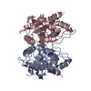
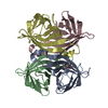
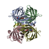
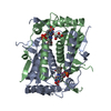
- PDB-1pts: CRYSTAL STRUCTURE AND LIGAND BINDING STUDIES OF A SCREENED PEPTID... -
+ Open data

- Basic information
| Entry | Database: PDB / ID: 1pts | ||||||
|---|---|---|---|---|---|---|---|
| Title | CRYSTAL STRUCTURE AND LIGAND BINDING STUDIES OF A SCREENED PEPTIDE COMPLEXED WITH STREPTAVIDIN | ||||||
 Components Components |
| ||||||
 Keywords Keywords | BIOTIN-BINDING PROTEIN/PEPTIDE /  GLYCOPROTEIN / BIOTIN-BINDING PROTEIN-PEPTIDE complex GLYCOPROTEIN / BIOTIN-BINDING PROTEIN-PEPTIDE complex | ||||||
| Function / homology |  Function and homology information Function and homology information | ||||||
| Biological species |  Streptomyces avidinii (bacteria) Streptomyces avidinii (bacteria) | ||||||
| Method | X-RAY DIFFRACTION / Resolution: 2 Å | ||||||
 Authors Authors | Weber, P.C. / Pantoliano, M.W. / Thompson, L.D. | ||||||
 Citation Citation | Journal: Biochemistry / Year: 1992 Title: Crystal structure and ligand-binding studies of a screened peptide complexed with streptavidin. Authors: Weber, P.C. / Pantoliano, M.W. / Thompson, L.D. #1: Journal: J.Am.Chem.Soc. / Year: 1992Title: Crystallographic and Thermodynamic Comparison of Natural and Synthetic Ligands Bound to Streptavidin Authors: Weber, P.C. / Wendoloski, J.J. / Pantoliano, M.W. / Salemme, F.R. #2: Journal: Science / Year: 1989Title: Structural Origins of High-Affinity Biotin Binding to Streptavidin Authors: Weber, P.C. / Ohlendorf, D.H. / Wendoloski, J.J. / Salemme, F.R. | ||||||
| History |
|
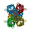
- Structure visualization
| Structure viewer | Molecule: MolmilJmol/JSmol |
|---|
- Downloads & links
-Download
| PDBx/mmCIF format | 1pts.cif.gz | 60.6 KB | Display | PDBx/mmCIF format |
|---|---|---|---|---|
| PDB format | pdb1pts.ent.gz | 44.5 KB | Display | PDB format |
| PDBx/mmJSON format | 1pts.json.gz | Tree view | PDBx/mmJSON format | |
| Others |  Other downloads Other downloads |
-Validation report
| Arichive directory | https://data.pdbj.org/pub/pdb/validation_reports/pt/1ptsftp://data.pdbj.org/pub/pdb/validation_reports/pt/1pts | HTTPS FTP |
|---|
-Related structure data
| Similar structure data |
|---|


-Links
 PDBj
PDBj- Assembly
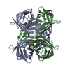
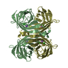
| Deposited unit | 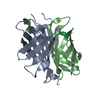
| ||||||||
|---|---|---|---|---|---|---|---|---|---|
| 1 | 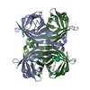
| ||||||||
| 2 | 
| ||||||||
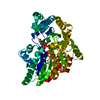
| Unit cell |
|
-Components
| #1: Protein | Mass: 12738.730 Da / Num. of mol.: 2 Source method: isolated from a genetically manipulated source Source: (gene. exp.) Streptomyces avidinii (bacteria) / References: UniProt: P22629#2: Protein/peptide | | Mass: 830.864 Da / Num. of mol.: 1 / Source method: obtained synthetically #3: Water | ChemComp-HOH / | Water Mass: 18.015 Da / Num. of mol.: 241 / Source method: isolated from a natural source / Formula: H2O Mass: 18.015 Da / Num. of mol.: 241 / Source method: isolated from a natural source / Formula: H2O |
|---|
-Experimental details
-Experiment
| Experiment | Method: X-RAY DIFFRACTION |
|---|
- Sample preparation
| Crystal | Density Matthews: 2.28 Å3/Da / Density % sol: 46.1 % |
|---|---|
| Crystal grow | *PLUS Method: unknown |
-Data collection
| Radiation | Scattering type: x-ray |
|---|---|
| Radiation wavelength | Relative weight: 1 |
| Reflection | *PLUS Highest resolution: 2 Å / Num. obs: 15804 / Rmerge(I) obs: 0.069 |
- Processing
| Software |
| ||||||||||||||||||||||||||||||||||||||||||||||||||||||||||||
|---|---|---|---|---|---|---|---|---|---|---|---|---|---|---|---|---|---|---|---|---|---|---|---|---|---|---|---|---|---|---|---|---|---|---|---|---|---|---|---|---|---|---|---|---|---|---|---|---|---|---|---|---|---|---|---|---|---|---|---|---|---|
| Refinement | Rfactor Rwork: 0.179 / Rfactor obs: 0.179 / Highest resolution: 2 Å | ||||||||||||||||||||||||||||||||||||||||||||||||||||||||||||
| Refinement step | Cycle: LAST / Highest resolution: 2 Å
| ||||||||||||||||||||||||||||||||||||||||||||||||||||||||||||
| Refine LS restraints |
| ||||||||||||||||||||||||||||||||||||||||||||||||||||||||||||
| Refinement | *PLUS Lowest resolution: 10 Å / Num. reflection obs: 15592 / Rfactor obs: 0.179 / Rfactor Rwork: 0.179 | ||||||||||||||||||||||||||||||||||||||||||||||||||||||||||||
| Solvent computation | *PLUS | ||||||||||||||||||||||||||||||||||||||||||||||||||||||||||||
| Displacement parameters | *PLUS | ||||||||||||||||||||||||||||||||||||||||||||||||||||||||||||
| Refine LS restraints | *PLUS Type: x_angle_d / Dev ideal: 0.039 |
