 Movie
Movie Controller

[English] 日本語
 Yorodumi
- PDB-1pne: CRYSTALLIZATION AND STRUCTURE DETERMINATION OF BOVINE PROFILIN AT... -
+ Open data

- Basic information
| Entry | Database: PDB / ID: 1pne | ||||||
|---|---|---|---|---|---|---|---|
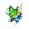
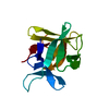
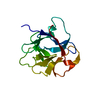
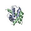
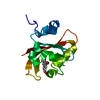
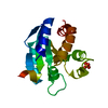
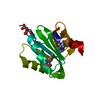
| Title | CRYSTALLIZATION AND STRUCTURE DETERMINATION OF BOVINE PROFILIN AT 2.0 ANGSTROMS RESOLUTION | ||||||
 Components Components | PROFILIN | ||||||
 Keywords Keywords | ACTIN BINDING PROTEIN | ||||||
| Function / homology |  Function and homology information Function and homology informationPCP/CE pathway / RHO GTPases Activate Formins / positive regulation of actin filament bundle assembly / regulation of actin filament polymerization / actin binding / actin cytoskeleton organization / cytoskeleton / cytoplasmSimilarity search - Function | ||||||
| Biological species |  Bos taurus (cattle) Bos taurus (cattle) | ||||||
| Method | X-RAY DIFFRACTION / Resolution: 2 Å | ||||||
 Authors Authors | Cedergren-Zeppezauer, E.S. / Goonesekere, N.C.W. / Rozycki, M.D. / Myslik, J.C. / Dauter, Z. / Lindberg, U. / Schutt, C.E. | ||||||
 Citation Citation | Journal: J.Mol.Biol. / Year: 1994 Title: Crystallization and structure determination of bovine profilin at 2.0 A resolution. Authors: Cedergren-Zeppezauer, E.S. / Goonesekere, N.C. / Rozycki, M.D. / Myslik, J.C. / Dauter, Z. / Lindberg, U. / Schutt, C.E. #1: Journal: Curr.Opin.Struct.Biol. / Year: 1994Title: Structural Aspects of Actin-Binding Proteins Authors: Rozycki, M.D. / Myslik, J.C. / Schutt, C.E. / Lindberg, U. #2: Journal: Nature / Year: 1993Title: The Structure of Crystalline Profilin-Beta-Actin Authors: Schutt, C.E. / Myslik, J.C. / Rozycki, M.D. / Goonesekere, N.C.W. / Lindberg, U. #3: Journal: FEBS Lett. / Year: 1993Title: Mutagenesis of Human Profilin Locates its Poly(L-Proline)-Binding Site to a Hydrophobic Patch of Aromatic Amino Acids Authors: Bjorkegren, C. / Rozycki, M. / Schutt, C.E. / Lindberg, U. / Karlsson, R. | ||||||
| History |
| ||||||
| Remark 700 | SHEET PROFILIN IS DESCRIBED IN JRNL REFERENCE AS HAVING A 6-MEMBERED BETA-SHEET WHICH SHARES A ...SHEET PROFILIN IS DESCRIBED IN JRNL REFERENCE AS HAVING A 6-MEMBERED BETA-SHEET WHICH SHARES A STRAND WITH A SECOND 2-MEMBERED SHEET. TO CONFORM WITH PDB GUIDELINES, THESE SHEETS ARE COMBINED INTO A SINGLE 7-MEMBERED SHEET (SP1) IN THIS FILE. STRAND NUMBERING ALSO DIFFERS FROM THE JRNL REFERENCE. RESIDUES TRP 31 AND ILE 73 ADOPT BETA-BULGE CONFORMATIONS. |
- Structure visualization
| Structure viewer | Molecule: MolmilJmol/JSmol |
|---|
- Downloads & links
-Download
| PDBx/mmCIF format | 1pne.cif.gz | 37.4 KB | Display | PDBx/mmCIF format |
|---|---|---|---|---|
| PDB format | pdb1pne.ent.gz | 28.1 KB | Display | PDB format |
| PDBx/mmJSON format | 1pne.json.gz | Tree view | PDBx/mmJSON format | |
| Others |  Other downloads Other downloads |
-Validation report
| Arichive directory | https://data.pdbj.org/pub/pdb/validation_reports/pn/1pneftp://data.pdbj.org/pub/pdb/validation_reports/pn/1pne | HTTPS FTP |
|---|
-Related structure data
| Similar structure data |
|---|


-Links
 PDBj
PDBj
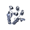
- Assembly
| Deposited unit | 
| ||||||||
|---|---|---|---|---|---|---|---|---|---|
| 1 |
| ||||||||
| Unit cell |
|
-Components
| #1: Protein | Mass: 14968.185 Da / Num. of mol.: 1 Source method: isolated from a genetically manipulated source Source: (gene. exp.) Bos taurus (cattle) / References: UniProt: P02584 |
|---|---|
| #2: Water | ChemComp-HOH / Water Mass: 18.015 Da / Num. of mol.: 127 / Source method: isolated from a natural source / Formula: H2O Mass: 18.015 Da / Num. of mol.: 127 / Source method: isolated from a natural source / Formula: H2O |
| Compound details | GAMMA-TURN AT RESIDUES 25 - 27 DOES NOT HAVE THE CANONICAL I TO I+2 HYDROGEN BOND, SINCE CARBONYL O ...GAMMA-TURN AT RESIDUES 25 - 27 DOES NOT HAVE THE CANONICAL I TO I+2 HYDROGEN BOND, SINCE CARBONYL O OF LYS 25 IS HYDROGEN BONDED TO WATER MOLECULE 27. WATER MOLECULE 27 IS BONDED TO WATER MOLECULE 28, WHICH IN TURN IS BOUND TO THE CARBONYL O OF SER 27. THE INVOLVEMEN |
| Sequence details | SEQUENCE IDENTIFIERS ARE NUMBERED 1 THROUGH 139 FOR STANDARD AMINO ACID RESIDUES. THE N-TERMINAL ...SEQUENCE IDENTIFIER |
-Experimental details
-Experiment
| Experiment | Method: X-RAY DIFFRACTION |
|---|
- Sample preparation
| Crystal | Density Matthews: 2.09 Å3/Da / Density % sol: 41.27 % | ||||||||||||||||||||||||||||||
|---|---|---|---|---|---|---|---|---|---|---|---|---|---|---|---|---|---|---|---|---|---|---|---|---|---|---|---|---|---|---|---|
| Crystal grow | *PLUS Temperature: 4 ℃ / pH: 7.5 / Method: vapor diffusion, sitting drop | ||||||||||||||||||||||||||||||
| Components of the solutions | *PLUS
|
-Data collection
| Detector | Date: 1993 |
|---|---|
| Radiation | Monochromatic (M) / Laue (L): M / Scattering type: x-ray |
| Radiation wavelength | Relative weight: 1 |
| Reflection | Num. obs: 8239 / % possible obs: 95.9 % / Observed criterion σ(I): 2 / Redundancy: 3.56 % / Rmerge(I) obs: 0.066 |
| Reflection | *PLUS Highest resolution: 2 Å / Num. measured all: 29297 / Rmerge(I) obs: 0.066 |
- Processing
| Software |
| ||||||||||||||||||||||||||||||||||||||||||||||||||||||||||||
|---|---|---|---|---|---|---|---|---|---|---|---|---|---|---|---|---|---|---|---|---|---|---|---|---|---|---|---|---|---|---|---|---|---|---|---|---|---|---|---|---|---|---|---|---|---|---|---|---|---|---|---|---|---|---|---|---|---|---|---|---|---|
| Refinement | Resolution: 2→6 Å / σ(F): 3 Details: RESIDUES ARE INCLUDED AT THE BEGINNING OF HELICES IF THEY PARTICIPATE IN HYDROGEN BONDING AND ARE "PARTIALLY" HELICAL, EVEN IF THE ENTIRE RESIDUE DOES NOT FIT HELICAL CRITERIA.
| ||||||||||||||||||||||||||||||||||||||||||||||||||||||||||||
| Refinement step | Cycle: LAST / Resolution: 2→6 Å
| ||||||||||||||||||||||||||||||||||||||||||||||||||||||||||||
| Refine LS restraints |
| ||||||||||||||||||||||||||||||||||||||||||||||||||||||||||||
| Software | *PLUS Name: X-PLOR / Classification: refinement | ||||||||||||||||||||||||||||||||||||||||||||||||||||||||||||
| Refine LS restraints | *PLUS
|
