 Movie
Movie Controller


+ Open data

- Basic information
| Entry | Database: PDB / ID: 1pex | ||||||
|---|---|---|---|---|---|---|---|
| Title | COLLAGENASE-3 (MMP-13) C-TERMINAL HEMOPEXIN-LIKE DOMAIN | ||||||
 Components Components | COLLAGENASE-3 | ||||||
 Keywords Keywords | METALLOPROTEASE / C-TERMINAL HEMOPEXIN-LIKE DOMAIN OF MATRIX-METALLOPROTEINASE | ||||||
| Function / homology |  Function and homology information Function and homology informationgrowth plate cartilage development / RUNX2 regulates genes involved in cell migration / endochondral ossification / Hydrolases; Acting on peptide bonds (peptidases); Metalloendopeptidases / bone morphogenesis / Assembly of collagen fibrils and other multimeric structures / bone mineralization / Activation of Matrix Metalloproteinases / response to amyloid-beta / Collagen degradation ...growth plate cartilage development / RUNX2 regulates genes involved in cell migration / endochondral ossification / Hydrolases; Acting on peptide bonds (peptidases); Metalloendopeptidases / bone morphogenesis / Assembly of collagen fibrils and other multimeric structures / bone mineralization / Activation of Matrix Metalloproteinases / response to amyloid-beta / Collagen degradation / collagen catabolic process / extracellular matrix disassembly / collagen binding / Degradation of the extracellular matrix / extracellular matrix organization / extracellular matrix / metalloendopeptidase activity / endopeptidase activity / serine-type endopeptidase activity / calcium ion binding / proteolysis / extracellular space / zinc ion binding / extracellular regionSimilarity search - Function | ||||||
| Biological species |  Homo sapiens (human) Homo sapiens (human) | ||||||
| Method | X-RAY DIFFRACTION / Resolution: 2.7 Å | ||||||
 Authors Authors | Gomis-Ruth, F.X. / Gohlke, U. / Betz, M. / Knauper, V. / Murphy, G. / Lopez-Otin, C. / Bode, W. | ||||||
 Citation Citation | Journal: J.Mol.Biol. / Year: 1996 Title: The helping hand of collagenase-3 (MMP-13): 2.7 A crystal structure of its C-terminal haemopexin-like domain. Authors: Gomis-Ruth, F.X. / Gohlke, U. / Betz, M. / Knauper, V. / Murphy, G. / Lopez-Otin, C. / Bode, W. | ||||||
| History |
|
- Structure visualization
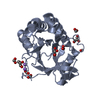
| Structure viewer | Molecule: MolmilJmol/JSmol |
|---|
- Downloads & links
-Download
| PDBx/mmCIF format | 1pex.cif.gz | 55.1 KB | Display | PDBx/mmCIF format |
|---|---|---|---|---|
| PDB format | pdb1pex.ent.gz | 39.4 KB | Display | PDB format |
| PDBx/mmJSON format | 1pex.json.gz | Tree view | PDBx/mmJSON format | |
| Others |  Other downloads Other downloads |
-Validation report
| Arichive directory | https://data.pdbj.org/pub/pdb/validation_reports/pe/1pexftp://data.pdbj.org/pub/pdb/validation_reports/pe/1pex | HTTPS FTP |
|---|
-Related structure data
| Similar structure data |
|---|


-Links
 PDBj
PDBj


- Assembly
| Deposited unit | 
| ||||||||
|---|---|---|---|---|---|---|---|---|---|
| 1 |
| ||||||||
| Unit cell |
|
-Components
| #1: Protein | / MMP-13 Mass: 24183.404 Da / Num. of mol.: 1 / Fragment: C-TERMINAL HEMOPEXIN-LIKE DOMAIN Source method: isolated from a genetically manipulated source Source: (gene. exp.) Homo sapiens (human) / Tissue: BREAST CANCER / Description: STABLY TRANSFECTED / Organ: BREAST / Cell (production host): 1PEX CELLSOrganelle (production host): PURIFIED FROM SERUM FREE CONDITIONED CELL CULTURE MEDIUM Production host:  Mus musculus (house mouse) Mus musculus (house mouse)References: UniProt: P45452, Hydrolases; Acting on peptide bonds (peptidases); Metalloendopeptidases | ||||
|---|---|---|---|---|---|
| #2: Chemical | ChemComp-SO4 / Sulfate  Mass: 96.063 Da / Num. of mol.: 1 / Source method: obtained synthetically / Formula: SO4 Mass: 96.063 Da / Num. of mol.: 1 / Source method: obtained synthetically / Formula: SO4 | ||||
| #3: Chemical | Chloride  Mass: 35.453 Da / Num. of mol.: 2 / Source method: obtained synthetically / Formula: Cl Mass: 35.453 Da / Num. of mol.: 2 / Source method: obtained synthetically / Formula: Cl#4: Chemical |   Mass: 40.078 Da / Num. of mol.: 2 / Source method: obtained synthetically / Formula: Ca Mass: 40.078 Da / Num. of mol.: 2 / Source method: obtained synthetically / Formula: Ca#5: Water | ChemComp-HOH / | Water Mass: 18.015 Da / Num. of mol.: 73 / Source method: isolated from a natural source / Formula: H2O Mass: 18.015 Da / Num. of mol.: 73 / Source method: isolated from a natural source / Formula: H2O |
-Experimental details
-Experiment
| Experiment | Method: X-RAY DIFFRACTION |
|---|
- Sample preparation
| Crystal | Density Matthews: 2.41 Å3/Da / Density % sol: 49 % | ||||||||||||||||||||||||||||||||||||||||||||||||
|---|---|---|---|---|---|---|---|---|---|---|---|---|---|---|---|---|---|---|---|---|---|---|---|---|---|---|---|---|---|---|---|---|---|---|---|---|---|---|---|---|---|---|---|---|---|---|---|---|---|
| Crystal | *PLUS | ||||||||||||||||||||||||||||||||||||||||||||||||
| Crystal grow | *PLUS Temperature: 20 ℃ / Method: vapor diffusion, hanging drop | ||||||||||||||||||||||||||||||||||||||||||||||||
| Components of the solutions | *PLUS
|
-Data collection
| Diffraction source | Wavelength: 1.5418 |
|---|---|
| Detector | Type: MARRESEARCH / Detector: IMAGE PLATE |
| Radiation | Monochromatic (M) / Laue (L): M / Scattering type: x-ray |
| Radiation wavelength | Wavelength: 1.5418 Å / Relative weight: 1 |
| Reflection | Resolution: 2.7→26.4 Å / Num. obs: 6858 / % possible obs: 95.7 % / Observed criterion σ(I): 3 / Rmerge(I) obs: 0.063 |
| Reflection | *PLUS Num. measured all: 24238 |
| Reflection shell | *PLUS Highest resolution: 2.7 Å / Lowest resolution: 2.82 Å / % possible obs: 94.9 % |
- Processing
| Software |
| ||||||||||||||||||||||||||||||||||||||||||||||||||||||||||||
|---|---|---|---|---|---|---|---|---|---|---|---|---|---|---|---|---|---|---|---|---|---|---|---|---|---|---|---|---|---|---|---|---|---|---|---|---|---|---|---|---|---|---|---|---|---|---|---|---|---|---|---|---|---|---|---|---|---|---|---|---|---|
| Refinement | Resolution: 2.7→6 Å /
| ||||||||||||||||||||||||||||||||||||||||||||||||||||||||||||
| Displacement parameters | Biso mean: 42.9 Å2 | ||||||||||||||||||||||||||||||||||||||||||||||||||||||||||||
| Refinement step | Cycle: LAST / Resolution: 2.7→6 Å
| ||||||||||||||||||||||||||||||||||||||||||||||||||||||||||||
| Refine LS restraints |
| ||||||||||||||||||||||||||||||||||||||||||||||||||||||||||||
| Software | *PLUS Name: X-PLOR / Classification: refinement | ||||||||||||||||||||||||||||||||||||||||||||||||||||||||||||
| Refinement | *PLUS Rfactor Rfree: 0.319 | ||||||||||||||||||||||||||||||||||||||||||||||||||||||||||||
| Solvent computation | *PLUS | ||||||||||||||||||||||||||||||||||||||||||||||||||||||||||||
| Displacement parameters | *PLUS |
