 Movie
Movie Controller

[English] 日本語
 Yorodumi
- PDB-1pe6: REFINED X-RAY STRUCTURE OF PAPAIN(DOT)E-64-C COMPLEX AT 2.1-ANGST... -
+ Open data

- Basic information
| Entry | Database: PDB / ID: 1pe6 | ||||||
|---|---|---|---|---|---|---|---|
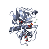
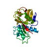
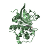
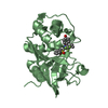
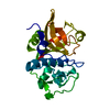
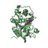
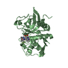
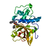
| Title | REFINED X-RAY STRUCTURE OF PAPAIN(DOT)E-64-C COMPLEX AT 2.1-ANGSTROMS RESOLUTION | ||||||
 Components Components | PAPAIN | ||||||
 Keywords Keywords | HYDROLASE (SULFHYDRYL PROTEINASE) | ||||||
| Function / homology |  Function and homology informationpapain / serpin family protein binding / cysteine-type peptidase activity / proteolysis Function and homology informationpapain / serpin family protein binding / cysteine-type peptidase activity / proteolysisSimilarity search - Function | ||||||
| Biological species |  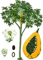 Carica papaya (papaya) Carica papaya (papaya) | ||||||
| Method | X-RAY DIFFRACTION / Resolution: 2.1 Å | ||||||
 Authors Authors | Yamamoto, D. / Matsumoto, K. / Ohishi, H. / Ishida, T. / Inoue, M. / Kitamura, K. / Mizuno, H. | ||||||
 Citation Citation | Journal: J.Biol.Chem. / Year: 1991 Title: Refined x-ray structure of papain.E-64-c complex at 2.1-A resolution. Authors: Yamamoto, D. / Matsumoto, K. / Ohishi, H. / Ishida, T. / Inoue, M. / Kitamura, K. / Mizuno, H. #1: Journal: FEBS Lett. / Year: 1989Title: Mode of Binding of E-64-C, a Potent Thiol Protease Inhibitor, to Papain as Determined by X-Ray Crystal Analysis of the Complex Authors: Matsumoto, K. / Yamamoto, D. / Ohishi, H. / Tomoo, K. / Ishida, T. / Inoue, M. / Sadatome, T. / Kitamura, K. / Mizuno, H. #2: Journal: FEBS Lett. / Year: 1990Title: The Importance of Val-157 Hydrophobic Interaction for Papain Inhibitory Activity of an Epoxysuccinyl Amino Acid Derivative; a Structure-Activity Relationship Based on the Crystal Structure of ...Title: The Importance of Val-157 Hydrophobic Interaction for Papain Inhibitory Activity of an Epoxysuccinyl Amino Acid Derivative; a Structure-Activity Relationship Based on the Crystal Structure of the Papain-E-64-C Complex Authors: Yamamoto, D. / Matsumoto, K. / Ohishi, H. / Ishida, T. / Inoue, M. / Kitamura, K. / Mizuno, H. | ||||||
| History |
| ||||||
| Remark 700 | SHEET THE SHEET STRUCTURE OF THIS MOLECULE IS BIFURCATED. IN ORDER TO REPRESENT THIS FEATURE IN THE ...SHEET THE SHEET STRUCTURE OF THIS MOLECULE IS BIFURCATED. IN ORDER TO REPRESENT THIS FEATURE IN THE SHEET RECORDS BELOW, TWO SHEETS ARE DEFINED. STRANDS 4, 5, 6 OF S1A ARE IDENTICAL TO STRANDS 2, 3, 4 OF SHEET S1B. |
- Structure visualization
| Structure viewer | Molecule: MolmilJmol/JSmol |
|---|
- Downloads & links
-Download
| PDBx/mmCIF format | 1pe6.cif.gz | 60.2 KB | Display | PDBx/mmCIF format |
|---|---|---|---|---|
| PDB format | pdb1pe6.ent.gz | 43.6 KB | Display | PDB format |
| PDBx/mmJSON format | 1pe6.json.gz | Tree view | PDBx/mmJSON format | |
| Others |  Other downloads Other downloads |
-Validation report
| Arichive directory | https://data.pdbj.org/pub/pdb/validation_reports/pe/1pe6ftp://data.pdbj.org/pub/pdb/validation_reports/pe/1pe6 | HTTPS FTP |
|---|
-Related structure data
| Similar structure data |
|---|


-Links
 PDBj
PDBj
- Assembly
| Deposited unit | 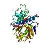
| ||||||||
|---|---|---|---|---|---|---|---|---|---|
| 1 |
| ||||||||
| Unit cell |
| ||||||||
| Atom site foot note | 1: RESIDUE PRO 152 IS A CIS PROLINE. / 2: SEE REMARK 6. |
-Components
| #1: Protein | Mass: 23449.346 Da / Num. of mol.: 1 Source method: isolated from a genetically manipulated source Source: (gene. exp.) Carica papaya (papaya) / References: UniProt: P00784, papain | ||||||
|---|---|---|---|---|---|---|---|
| #2: Chemical | ChemComp-E6C /   Mass: 316.393 Da / Num. of mol.: 1 / Source method: obtained synthetically / Formula: C15H28N2O5 Mass: 316.393 Da / Num. of mol.: 1 / Source method: obtained synthetically / Formula: C15H28N2O5 | ||||||
| #3: Chemical | ChemComp-MOH / Methanol  Mass: 32.042 Da / Num. of mol.: 14 / Source method: obtained synthetically / Formula: CH4O Mass: 32.042 Da / Num. of mol.: 14 / Source method: obtained synthetically / Formula: CH4O#4: Water | ChemComp-HOH / | Water Mass: 18.015 Da / Num. of mol.: 181 / Source method: isolated from a natural source / Formula: H2O Mass: 18.015 Da / Num. of mol.: 181 / Source method: isolated from a natural source / Formula: H2OCompound details | THE SG ATOM OF CYS 25, THE CATALYTIC CENTER OF PAPAIN, COVALENTLY BONDS TO THE C2 ATOM OF E-64-C, ...THE SG ATOM OF CYS 25, THE CATALYTIC CENTER OF PAPAIN, COVALENTLY | Nonpolymer details | E6C IS (2S,3S)-3-(1-(N-(3-METHYLBUTYL)AMINO)- LEUCYLCARBOXYL)OXIRANE-2-CARBOXYLATE. THIS COMPOUND ...E6C IS (2S,3S)-3-(1-(N-(3-METHYLBUTY | |
-Experimental details
-Experiment
| Experiment | Method: X-RAY DIFFRACTION |
|---|
- Sample preparation
| Crystal | Density Matthews: 2.18 Å3/Da / Density % sol: 43.65 % | ||||||||||||||||||||||||||||||
|---|---|---|---|---|---|---|---|---|---|---|---|---|---|---|---|---|---|---|---|---|---|---|---|---|---|---|---|---|---|---|---|
| Crystal grow | *PLUS Temperature: 20 ℃ / Method: vapor diffusion, hanging drop | ||||||||||||||||||||||||||||||
| Components of the solutions | *PLUS
|
-Data collection
| Radiation | Scattering type: x-ray |
|---|---|
| Radiation wavelength | Relative weight: 1 |
| Reflection | *PLUS Highest resolution: 2.1 Å / Num. obs: 12794 / Rmerge(I) obs: 0.027 / Biso Wilson estimate: 11.1 Å2 |
- Processing
| Software | Name: PROLSQ / Classification: refinement | ||||||||||||||||||||||||||||||||||||||||||||||||||||||||||||||||||||||||||||||||||||
|---|---|---|---|---|---|---|---|---|---|---|---|---|---|---|---|---|---|---|---|---|---|---|---|---|---|---|---|---|---|---|---|---|---|---|---|---|---|---|---|---|---|---|---|---|---|---|---|---|---|---|---|---|---|---|---|---|---|---|---|---|---|---|---|---|---|---|---|---|---|---|---|---|---|---|---|---|---|---|---|---|---|---|---|---|---|
| Refinement | Resolution: 2.1→6 Å /
| ||||||||||||||||||||||||||||||||||||||||||||||||||||||||||||||||||||||||||||||||||||
| Refinement step | Cycle: LAST / Resolution: 2.1→6 Å
| ||||||||||||||||||||||||||||||||||||||||||||||||||||||||||||||||||||||||||||||||||||
| Refine LS restraints |
|
