 Movie
Movie Controller

[English] 日本語
 Yorodumi
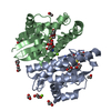
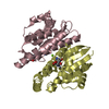
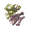
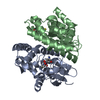
- PDB-1pd2: CRYSTAL STRUCTURE OF HEMATOPOIETIC PROSTAGLANDIN D SYNTHASE COMPL... -
+ Open data

- Basic information
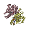
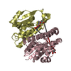
| Entry | Database: PDB / ID: 1pd2 | ||||||
|---|---|---|---|---|---|---|---|
| Title | CRYSTAL STRUCTURE OF HEMATOPOIETIC PROSTAGLANDIN D SYNTHASE COMPLEX WITH GLUTATHIONE | ||||||
 Components Components | HEMATOPOIETIC PROSTAGLANDIN D SYNTHASE | ||||||
 Keywords Keywords |  LIGASE / HEMATOPOIETIC PROSTAGLANDIN D SYNTHASE / PGDS / GST / SIGMA-CLASS GST LIGASE / HEMATOPOIETIC PROSTAGLANDIN D SYNTHASE / PGDS / GST / SIGMA-CLASS GST | ||||||
| Function / homology |  Function and homology information Function and homology informationSynthesis of Prostaglandins (PG) and Thromboxanes (TX) / Glutathione conjugation / prostaglandin-D synthase / prostaglandin-D synthase activity / negative regulation of male germ cell proliferation / prostaglandin biosynthetic process / prostaglandin metabolic process / glutathione transferase / glutathione transferase activity / calcium ion binding ...Synthesis of Prostaglandins (PG) and Thromboxanes (TX) / Glutathione conjugation / prostaglandin-D synthase / prostaglandin-D synthase activity / negative regulation of male germ cell proliferation / prostaglandin biosynthetic process / prostaglandin metabolic process / glutathione transferase / glutathione transferase activity / calcium ion binding / magnesium ion binding / protein homodimerization activity / cytoplasmSimilarity search - Function | ||||||
| Biological species |  Rattus norvegicus (Norway rat) Rattus norvegicus (Norway rat) | ||||||
| Method | X-RAY DIFFRACTION / SYNCHROTRON / MIRAS / Resolution: 2.3 Å | ||||||
 Authors Authors | Miyano, M. / Ago, H. | ||||||
 Citation Citation | Journal: Cell(Cambridge,Mass.) / Year: 1997 Title: Cloning and crystal structure of hematopoietic prostaglandin D synthase. Authors: Kanaoka, Y. / Ago, H. / Inagaki, E. / Nanayama, T. / Miyano, M. / Kikuno, R. / Fujii, Y. / Eguchi, N. / Toh, H. / Urade, Y. / Hayaishi, O. #1: Journal: Cell(Cambridge,Mass.) / Year: 1999Title: Erratum. Cloning and Crystal Structure of Hematopoietic Prostaglandin D Synthase Authors: Kanaoka, Y. / Ago, H. / Inagaki, E. / Nanayama, T. / Miyano, M. / Kikuno, R. / Fujii, Y. / Eguchi, N. / Toh, H. / Urade, Y. / Hayaishi, O. | ||||||
| History |
|
- Structure visualization
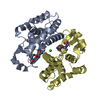
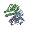
| Structure viewer | Molecule: MolmilJmol/JSmol |
|---|
- Downloads & links
-Download
| PDBx/mmCIF format | 1pd2.cif.gz | 113.1 KB | Display | PDBx/mmCIF format |
|---|---|---|---|---|
| PDB format | pdb1pd2.ent.gz | 89.4 KB | Display | PDB format |
| PDBx/mmJSON format | 1pd2.json.gz | Tree view | PDBx/mmJSON format | |
| Others |  Other downloads Other downloads |
-Validation report
| Arichive directory | https://data.pdbj.org/pub/pdb/validation_reports/pd/1pd2ftp://data.pdbj.org/pub/pdb/validation_reports/pd/1pd2 | HTTPS FTP |
|---|
-Related structure data
| Similar structure data |
|---|


-Links
 PDBj
PDBj

- Assembly
| Deposited unit | 
| ||||||||
|---|---|---|---|---|---|---|---|---|---|
| 1 |
| ||||||||
| Unit cell |
| ||||||||
| Noncrystallographic symmetry (NCS) | NCS oper: (Code: given Matrix: (-0.969084, 0.134643, 0.206756), Vector : |
-Components
| #1: Protein | Mass: 23323.943 Da / Num. of mol.: 2 Source method: isolated from a genetically manipulated source Source: (gene. exp.) Rattus norvegicus (Norway rat) / Cellular location: CYTOPLASM / Organ: SPLEEN / Species (production host): Escherichia coli / Cellular location (production host): CYTOPLASMIC / Gene (production host): DE3 / Production host:  Escherichia coli BL21(DE3) (bacteria) / Strain (production host): BL21 / Variant (production host): DE3 / References: UniProt: O35543, prostaglandin-D synthase Escherichia coli BL21(DE3) (bacteria) / Strain (production host): BL21 / Variant (production host): DE3 / References: UniProt: O35543, prostaglandin-D synthase#2: Chemical | Glutathione  Mass: 307.323 Da / Num. of mol.: 2 / Source method: obtained synthetically / Formula: C10H17N3O6S Mass: 307.323 Da / Num. of mol.: 2 / Source method: obtained synthetically / Formula: C10H17N3O6S#3: Water | ChemComp-HOH / | Water Mass: 18.015 Da / Num. of mol.: 99 / Source method: isolated from a natural source / Formula: H2O Mass: 18.015 Da / Num. of mol.: 99 / Source method: isolated from a natural source / Formula: H2O |
|---|
-Experimental details
-Experiment
| Experiment | Method: X-RAY DIFFRACTION / Number of used crystals: 1 |
|---|
- Sample preparation
| Crystal | Density Matthews: 2.2 Å3/Da / Density % sol: 46.62 % | ||||||||||||||||||||||||||||||||||||||||||||||||||||||
|---|---|---|---|---|---|---|---|---|---|---|---|---|---|---|---|---|---|---|---|---|---|---|---|---|---|---|---|---|---|---|---|---|---|---|---|---|---|---|---|---|---|---|---|---|---|---|---|---|---|---|---|---|---|---|---|
| Crystal grow | Details: VAPOR DIFFUSION METHOD RESERVOIR SOLUTION: 18% (W/V) PEG 6K, 100 MM AMMONIUM CITRATE 5MM CACL2, 10MM DTT, 2% (V/V) 1,4-DIOXANE PROTEIN SOLUTION: 7.3 MG/ML PROTEIN (50 MM TRIS-HCL [PH 7.5]) ...Details: VAPOR DIFFUSION METHOD RESERVOIR SOLUTION: 18% (W/V) PEG 6K, 100 MM AMMONIUM CITRATE 5MM CACL2, 10MM DTT, 2% (V/V) 1,4-DIOXANE PROTEIN SOLUTION: 7.3 MG/ML PROTEIN (50 MM TRIS-HCL [PH 7.5]) EACH 4 UL WAS MIXED FOR DROPLET | ||||||||||||||||||||||||||||||||||||||||||||||||||||||
| Components of the solutions |
| ||||||||||||||||||||||||||||||||||||||||||||||||||||||
| Crystal grow | *PLUS Temperature: 22.5 ℃ / pH: 7.5 / Method: vapor diffusion, hanging drop | ||||||||||||||||||||||||||||||||||||||||||||||||||||||
| Components of the solutions | *PLUS
|
-Data collection
| Diffraction | Mean temperature: 298 K |
|---|---|
| Diffraction source | Source: SYNCHROTRON / Site: Photon Factory  / Beamline: BL-6A / Wavelength: 1 / Beamline: BL-6A / Wavelength: 1 |
| Detector | Type: RIGAKU / Detector: IMAGE PLATE |
| Radiation | Protocol: SINGLE WAVELENGTH / Monochromatic (M) / Laue (L): M / Scattering type: x-ray |
| Radiation wavelength | Wavelength: 1 Å / Relative weight: 1 |
| Reflection | Resolution: 2.3→40 Å / Num. obs: 77899 / % possible obs: 95.1 % / Redundancy: 4 % / Rmerge(I) obs: 0.085 |
- Processing
| Software |
| ||||||||||||||||||||||||||||||||||||||||||||||||||||||||||||
|---|---|---|---|---|---|---|---|---|---|---|---|---|---|---|---|---|---|---|---|---|---|---|---|---|---|---|---|---|---|---|---|---|---|---|---|---|---|---|---|---|---|---|---|---|---|---|---|---|---|---|---|---|---|---|---|---|---|---|---|---|---|
| Refinement | Method to determine structure: MIRAS / Resolution: 2.3→6 Å / Cross valid method: PROCHECK / σ(F): 2
| ||||||||||||||||||||||||||||||||||||||||||||||||||||||||||||
| Refine analyze | Luzzati coordinate error obs: 0.2 Å | ||||||||||||||||||||||||||||||||||||||||||||||||||||||||||||
| Refinement step | Cycle: LAST / Resolution: 2.3→6 Å
| ||||||||||||||||||||||||||||||||||||||||||||||||||||||||||||
| Refine LS restraints |
| ||||||||||||||||||||||||||||||||||||||||||||||||||||||||||||
| Refine LS restraints NCS | NCS model details: RESTRAINTS | ||||||||||||||||||||||||||||||||||||||||||||||||||||||||||||
| LS refinement shell | Resolution: 2.3→2.4 Å / Total num. of bins used: 8
| ||||||||||||||||||||||||||||||||||||||||||||||||||||||||||||
| Software | *PLUS Name: X-PLOR / Version: 3.1 / Classification: refinement | ||||||||||||||||||||||||||||||||||||||||||||||||||||||||||||
| Refinement | *PLUS Highest resolution: 2.3 Å / Lowest resolution: 6 Å / σ(F): 2 / % reflection Rfree: 10 % / Rfactor obs: 0.204 | ||||||||||||||||||||||||||||||||||||||||||||||||||||||||||||
| Solvent computation | *PLUS | ||||||||||||||||||||||||||||||||||||||||||||||||||||||||||||
| Displacement parameters | *PLUS | ||||||||||||||||||||||||||||||||||||||||||||||||||||||||||||
| LS refinement shell | *PLUS Highest resolution: 2.3 Å / Lowest resolution: 2.4 Å / % reflection Rfree: 5 % |
