 Movie
Movie Controller

[English] 日本語
 Yorodumi
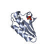
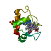
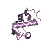
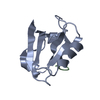
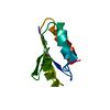
- PDB-1pch: STRUCTURAL EVIDENCE FOR THE EVOLUTIONARY DIVERGENCE OF MYCOPLASMA... -
+ Open data

- Basic information
| Entry | Database: PDB / ID: 1pch | ||||||
|---|---|---|---|---|---|---|---|
| Title | STRUCTURAL EVIDENCE FOR THE EVOLUTIONARY DIVERGENCE OF MYCOPLASMA FROM GRAM-POSITIVE BACTERIA: THE HISTIDINE-CONTAINING PHOSPHOCARRIER PROTEIN | ||||||
 Components Components | PHOSPHOCARRIER PROTEIN | ||||||
 Keywords Keywords |  PHOSPHOTRANSFERASE PHOSPHOTRANSFERASE | ||||||
| Function / homology |  Function and homology information Function and homology informationphosphoenolpyruvate-dependent sugar phosphotransferase system / cytoplasmSimilarity search - Function | ||||||
| Biological species |  Mycoplasma capricolum (bacteria) Mycoplasma capricolum (bacteria) | ||||||
| Method | X-RAY DIFFRACTION / Resolution: 1.8 Å | ||||||
 Authors Authors | Pieper, U. / Herzberg, O. | ||||||
 Citation Citation | Journal: Structure / Year: 1995 Title: Structural evidence for the evolutionary divergence of mycoplasma from gram-positive bacteria: the histidine-containing phosphocarrier protein. Authors: Pieper, U. / Kapadia, G. / Zhu, P.P. / Peterkofsky, A. / Herzberg, O. #1: Journal: Structure / Year: 1994Title: Refined Structures of the Active Ser83->Cys and Impaired Ser46->Asp Histidine Containing Phosphocarrier Proteins Authors: Liao, D.-I. / Herzberg, O. #2: Journal: Proc.Natl.Acad.Sci.USA / Year: 1992Title: Structure of the Histidine-Containing Phosphocarrier Protein Hpr from Bacillus Subtilis at 2.0-Angstroms Resolution Authors: Herzberg, O. / Reddy, P. / Sutrina, S. / Saier Junior, M.H. / Reizer, J. / Kapadia, G. | ||||||
| History |
|
- Structure visualization
| Structure viewer | Molecule: MolmilJmol/JSmol |
|---|
- Downloads & links
-Download
| PDBx/mmCIF format | 1pch.cif.gz | 28.5 KB | Display | PDBx/mmCIF format |
|---|---|---|---|---|
| PDB format | pdb1pch.ent.gz | 18.5 KB | Display | PDB format |
| PDBx/mmJSON format | 1pch.json.gz | Tree view | PDBx/mmJSON format | |
| Others |  Other downloads Other downloads |
-Validation report
| Arichive directory | https://data.pdbj.org/pub/pdb/validation_reports/pc/1pchftp://data.pdbj.org/pub/pdb/validation_reports/pc/1pch | HTTPS FTP |
|---|
-Related structure data
| Similar structure data |
|---|


-Links
 PDBj
PDBj- Assembly
| Deposited unit | 
| ||||||||
|---|---|---|---|---|---|---|---|---|---|
| 1 |
| ||||||||
| Unit cell |
|
-Components
| #1: Protein | Mass: 9296.718 Da / Num. of mol.: 1 Source method: isolated from a genetically manipulated source Source: (gene. exp.) Mycoplasma capricolum (bacteria) / Gene: PTSH / Plasmid: PRE1 / Gene (production host): PTSH / Production host: Escherichia coli (E. coli) / References: UniProt: P45611 |
|---|---|
| #2: Chemical | ChemComp-SO4 / Sulfate  Mass: 96.063 Da / Num. of mol.: 1 / Source method: obtained synthetically / Formula: SO4 Mass: 96.063 Da / Num. of mol.: 1 / Source method: obtained synthetically / Formula: SO4 |
| #3: Water | ChemComp-HOH / Water Mass: 18.015 Da / Num. of mol.: 60 / Source method: isolated from a natural source / Formula: H2O Mass: 18.015 Da / Num. of mol.: 60 / Source method: isolated from a natural source / Formula: H2O |
| Sequence details | MET 1 WAS PROCESSED DURING THE EXPRESSION |
-Experimental details
-Experiment
| Experiment | Method: X-RAY DIFFRACTION / Number of used crystals: 1 |
|---|
- Sample preparation
| Crystal | Density Matthews: 1.91 Å3/Da / Density % sol: 35.75 % | ||||||||||||||||||||||||||||||
|---|---|---|---|---|---|---|---|---|---|---|---|---|---|---|---|---|---|---|---|---|---|---|---|---|---|---|---|---|---|---|---|
| Crystal grow | *PLUS pH: 7.5 / Method: vapor diffusion, hanging drop | ||||||||||||||||||||||||||||||
| Components of the solutions | *PLUS
|
-Data collection
| Diffraction source | Wavelength: 1.5418 Å |
|---|---|
| Detector | Type: SIEMENS / Detector: AREA DETECTOR / Date: May 17, 1994 |
| Radiation | Monochromatic (M) / Laue (L): M / Scattering type: x-ray |
| Radiation wavelength | Wavelength: 1.5418 Å / Relative weight: 1 |
| Reflection | Resolution: 1.8→15 Å / Num. obs: 6562 / % possible obs: 94 % / Observed criterion σ(I): 0 / Redundancy: 2.8 % / Rmerge(I) obs: 0.045 |
| Reflection | *PLUS Num. all: 7044 / Num. measured all: 18520 / Rmerge(I) obs: 0.045 |
- Processing
| Software |
| ||||||||||||||||||||||||||||||||||||||||||||||||||||||||||||
|---|---|---|---|---|---|---|---|---|---|---|---|---|---|---|---|---|---|---|---|---|---|---|---|---|---|---|---|---|---|---|---|---|---|---|---|---|---|---|---|---|---|---|---|---|---|---|---|---|---|---|---|---|---|---|---|---|---|---|---|---|---|
| Refinement | Resolution: 1.8→15 Å / σ(F): 0 Details: TWO ALTERNATE CONFORMATIONS HAVE BEEN REFINED FOR THE SIDE CHAIN OF MET 51 WITH OCCUPANCIES OF 0.6 AND 0.4, RESPECTIVELY.
| ||||||||||||||||||||||||||||||||||||||||||||||||||||||||||||
| Refinement step | Cycle: LAST / Resolution: 1.8→15 Å
| ||||||||||||||||||||||||||||||||||||||||||||||||||||||||||||
| Refine LS restraints |
| ||||||||||||||||||||||||||||||||||||||||||||||||||||||||||||
| Refinement | *PLUS | ||||||||||||||||||||||||||||||||||||||||||||||||||||||||||||
| Solvent computation | *PLUS | ||||||||||||||||||||||||||||||||||||||||||||||||||||||||||||
| Displacement parameters | *PLUS Biso mean: 23.7 Å2 | ||||||||||||||||||||||||||||||||||||||||||||||||||||||||||||
| Refine LS restraints | *PLUS
|
