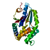
 Movie
Movie Controller


+ Open data

- Basic information
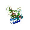
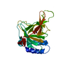
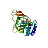
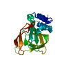
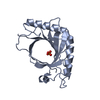
| Entry | Database: PDB / ID: 1pbw | ||||||
|---|---|---|---|---|---|---|---|
| Title | STRUCTURE OF BCR-HOMOLOGY (BH) DOMAIN | ||||||
 Components Components | PHOSPHATIDYLINOSITOL 3-KINASE Phosphoinositide 3-kinase Phosphoinositide 3-kinase | ||||||
 Keywords Keywords | PHOSPHOTRANSFERASE / TPASE ACTIVATING PROTEIN / GAP / CDC42 / PHOSPHOINOSITIDE 3-KINASE / SH3 DOMAIN / SH2 DOMAIN / SIGNAL TRANSDUCTION | ||||||
| Function / homology |  Function and homology information Function and homology informationperinuclear endoplasmic reticulum membrane / regulation of toll-like receptor 4 signaling pathway / phosphatidylinositol kinase activity / phosphatidylinositol 3-kinase regulator activity / positive regulation of focal adhesion disassembly / IRS-mediated signalling / phosphatidylinositol 3-kinase activator activity / interleukin-18-mediated signaling pathway / PI3K events in ERBB4 signaling / myeloid leukocyte migration ...perinuclear endoplasmic reticulum membrane / regulation of toll-like receptor 4 signaling pathway / phosphatidylinositol kinase activity / phosphatidylinositol 3-kinase regulator activity / positive regulation of focal adhesion disassembly / IRS-mediated signalling / phosphatidylinositol 3-kinase activator activity / interleukin-18-mediated signaling pathway / PI3K events in ERBB4 signaling / myeloid leukocyte migration / 1-phosphatidylinositol-3-kinase regulator activity / phosphatidylinositol 3-kinase regulatory subunit binding / neurotrophin TRKA receptor binding / Activated NTRK2 signals through PI3K / cis-Golgi network / Activated NTRK3 signals through PI3K / ErbB-3 class receptor binding / RHOF GTPase cycle / kinase activator activity / transmembrane receptor protein tyrosine kinase adaptor activity / RHOD GTPase cycle / Signaling by cytosolic FGFR1 fusion mutants / positive regulation of endoplasmic reticulum unfolded protein response / enzyme-substrate adaptor activity / phosphatidylinositol 3-kinase complex, class IA / phosphatidylinositol 3-kinase complex / Nephrin family interactions / RND1 GTPase cycle / Costimulation by the CD28 family / RND2 GTPase cycle / MET activates PI3K/AKT signaling / PI3K/AKT activation / RND3 GTPase cycle / positive regulation of leukocyte migration / positive regulation of filopodium assembly / negative regulation of stress fiber assembly / growth hormone receptor signaling pathway / insulin binding / RHOV GTPase cycle / RHOB GTPase cycle / negative regulation of cell-matrix adhesion / Signaling by ALK / GP1b-IX-V activation signalling / PI-3K cascade:FGFR3 / Erythropoietin activates Phosphoinositide-3-kinase (PI3K) / PI-3K cascade:FGFR2 / RHOJ GTPase cycle / PI-3K cascade:FGFR4 / RHOC GTPase cycle / PI-3K cascade:FGFR1 / negative regulation of osteoclast differentiation / intracellular glucose homeostasis / phosphatidylinositol phosphate biosynthetic process / CD28 dependent PI3K/Akt signaling / Synthesis of PIPs at the plasma membrane / CDC42 GTPase cycle / RHOU GTPase cycle / PI3K events in ERBB2 signaling / RHOG GTPase cycle / T cell differentiation / RET signaling / extrinsic apoptotic signaling pathway via death domain receptors / insulin receptor substrate binding / Interleukin-3, Interleukin-5 and GM-CSF signaling / PI3K Cascade / RHOA GTPase cycle / RAC2 GTPase cycle / RAC3 GTPase cycle / Role of phospholipids in phagocytosis / GAB1 signalosome / Role of LAT2/NTAL/LAB on calcium mobilization / Interleukin receptor SHC signaling / positive regulation of lamellipodium assembly / phosphatidylinositol 3-kinase binding / Signaling by PDGFRA transmembrane, juxtamembrane and kinase domain mutants / Signaling by PDGFRA extracellular domain mutants / Signaling by FGFR4 in disease / Signaling by FLT3 ITD and TKD mutants / Signaling by FGFR3 in disease / GPVI-mediated activation cascade / Tie2 Signaling / Signaling by FGFR2 in disease / RAC1 GTPase cycle / insulin-like growth factor receptor binding / Signaling by FLT3 fusion proteins / FLT3 Signaling / Signaling by FGFR1 in disease / Interleukin-7 signaling / phosphotyrosine residue binding / response to endoplasmic reticulum stress / Downstream signal transduction / substrate adhesion-dependent cell spreading / B cell differentiation / osteoclast differentiation / positive regulation of RNA splicing / insulin-like growth factor receptor signaling pathway / Signaling by phosphorylated juxtamembrane, extracellular and kinase domain KIT mutants / phosphatidylinositol 3-kinase/protein kinase B signal transduction / Antigen activates B Cell Receptor (BCR) leading to generation of second messengers / Regulation of signaling by CBLSimilarity search - Function | ||||||
| Biological species |  Homo sapiens (human) Homo sapiens (human) | ||||||
| Method | X-RAY DIFFRACTION / Resolution: 2 Å | ||||||
 Authors Authors | Musacchio, A. / Cantley, L.C. / Harrison, S.C. | ||||||
 Citation Citation | Journal: Proc.Natl.Acad.Sci.USA / Year: 1996 Title: Crystal structure of the breakpoint cluster region-homology domain from phosphoinositide 3-kinase p85 alpha subunit. Authors: Musacchio, A. / Cantley, L.C. / Harrison, S.C. #1: Journal: Cell(Cambridge,Mass.) / Year: 1991Title: Cloning of Pi3 Kinase-Associated P85 Utilizing a Novel Method for Expression/Cloning of Target Proteins for Receptor Tyrosine Kinases Authors: Skolnik, E.Y. / Margolis, B. / Mohammadi, M. / Lowenstein, E. / Fischer, R. / Drepps, A. / Ullrich, A. / Schlessinger, J. | ||||||
| History |
|
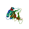
- Structure visualization
| Structure viewer | Molecule: MolmilJmol/JSmol |
|---|
- Downloads & links
-Download
| PDBx/mmCIF format | 1pbw.cif.gz | 93.5 KB | Display | PDBx/mmCIF format |
|---|---|---|---|---|
| PDB format | pdb1pbw.ent.gz | 71.4 KB | Display | PDB format |
| PDBx/mmJSON format | 1pbw.json.gz | Tree view | PDBx/mmJSON format | |
| Others |  Other downloads Other downloads |
-Validation report
| Arichive directory | https://data.pdbj.org/pub/pdb/validation_reports/pb/1pbwftp://data.pdbj.org/pub/pdb/validation_reports/pb/1pbw | HTTPS FTP |
|---|
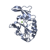
-Related structure data
| Similar structure data |
|---|

-Links
 PDBj
PDBj


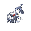
- Assembly
| Deposited unit | 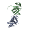
| ||||||||
|---|---|---|---|---|---|---|---|---|---|
| 1 | 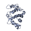
| ||||||||
| 2 | 
| ||||||||
| Unit cell |
| ||||||||
| Noncrystallographic symmetry (NCS) | NCS oper: (Code: given Matrix: (-0.055, 0.719, 0.413), Vector : |
-Components
| #1: Protein | Phosphoinositide 3-kinase / RHOGAP DOMAIN Mass: 24304.002 Da / Num. of mol.: 2 / Fragment: P85 ALPHA SUBUNIT BCR-HOMOLOGY DOMAIN Source method: isolated from a genetically manipulated source Source: (gene. exp.) Homo sapiens (human) / Description: T7-BASED EXPRESSION SYSTEM / Plasmid: PBAT4 / Production host:  Escherichia coli (E. coli) / References: UniProt: P27986 Escherichia coli (E. coli) / References: UniProt: P27986#2: Water | ChemComp-HOH / | Water Mass: 18.015 Da / Num. of mol.: 267 / Source method: isolated from a natural source / Formula: H2O Mass: 18.015 Da / Num. of mol.: 267 / Source method: isolated from a natural source / Formula: H2O |
|---|
-Experimental details
-Experiment
| Experiment | Method: X-RAY DIFFRACTION |
|---|
- Sample preparation
| Crystal | Density Matthews: 2.5 Å3/Da / Density % sol: 40 % | |||||||||||||||
|---|---|---|---|---|---|---|---|---|---|---|---|---|---|---|---|---|
| Crystal grow | *PLUS pH: 5 / Method: vapor diffusion, hanging drop | |||||||||||||||
| Components of the solutions | *PLUS
|
-Data collection
| Diffraction source | Wavelength: 1.5418 |
|---|---|
| Detector | Type: MARRESEARCH / Detector: IMAGE PLATE / Date: Apr 6, 1996 |
| Radiation | Monochromatic (M) / Laue (L): M / Scattering type: x-ray |
| Radiation wavelength | Wavelength: 1.5418 Å / Relative weight: 1 |
| Reflection | Num. obs: 30040 / % possible obs: 95 % / Redundancy: 2.54 % / Rmerge(I) obs: 0.057 |
| Reflection | *PLUS Highest resolution: 2 Å / Num. measured all: 124328 |
| Reflection shell | *PLUS % possible obs: 84 % / Rmerge(I) obs: 0.193 / Mean I/σ(I) obs: 5.4 |
- Processing
| Software |
| ||||||||||||||||||||||||||||||||||||||||||||||||||||||||||||
|---|---|---|---|---|---|---|---|---|---|---|---|---|---|---|---|---|---|---|---|---|---|---|---|---|---|---|---|---|---|---|---|---|---|---|---|---|---|---|---|---|---|---|---|---|---|---|---|---|---|---|---|---|---|---|---|---|---|---|---|---|---|
| Refinement | Resolution: 2→8 Å / σ(F): 0
| ||||||||||||||||||||||||||||||||||||||||||||||||||||||||||||
| Refinement step | Cycle: LAST / Resolution: 2→8 Å
| ||||||||||||||||||||||||||||||||||||||||||||||||||||||||||||
| Refine LS restraints |
| ||||||||||||||||||||||||||||||||||||||||||||||||||||||||||||
| Software | *PLUS Name: X-PLOR / Classification: refinement | ||||||||||||||||||||||||||||||||||||||||||||||||||||||||||||
| Refinement | *PLUS | ||||||||||||||||||||||||||||||||||||||||||||||||||||||||||||
| Solvent computation | *PLUS | ||||||||||||||||||||||||||||||||||||||||||||||||||||||||||||
| Displacement parameters | *PLUS |
