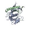
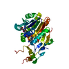
[English] 日本語
Yorodumi
- PDB-1pb8: CRYSTAL STRUCTURE OF THE NR1 LIGAND BINDING CORE IN COMPLEX WITH ... -

+
Open data


ID or keywords:

Loading...

-
Basic information

Entry
Database: PDB / ID: 1pb8
TitleCRYSTAL STRUCTURE OF THE NR1 LIGAND BINDING CORE IN COMPLEX WITH D-SERINE AT 1.45 ANGSTROMS RESOLUTION
ComponentsN-methyl-D-aspartate Receptor Subunit 1NMDA receptor
KeywordsLIGAND BINDING PROTEIN / Ligand binding receptor / rat / NR1
Function / homology
Function and homology information


pons maturation / regulation of cell communication / positive regulation of Schwann cell migration / EPHB-mediated forward signaling / Assembly and cell surface presentation of NMDA receptors / olfactory learning / conditioned taste aversion / protein localization to postsynaptic membrane / dendritic branch / regulation of respiratory gaseous exchange ...pons maturation / regulation of cell communication / positive regulation of Schwann cell migration / EPHB-mediated forward signaling / Assembly and cell surface presentation of NMDA receptors / olfactory learning / conditioned taste aversion / protein localization to postsynaptic membrane / dendritic branch / regulation of respiratory gaseous exchange / propylene metabolic process / response to glycine / voltage-gated monoatomic cation channel activity / response to morphine / glutamate-gated calcium ion channel activity / regulation of monoatomic cation transmembrane transport / NMDA glutamate receptor activity / NMDA selective glutamate receptor complex / RAF/MAP kinase cascade / Synaptic adhesion-like molecules / parallel fiber to Purkinje cell synapse / calcium ion transmembrane import into cytosol / protein heterotetramerization / glutamate binding / positive regulation of reactive oxygen species biosynthetic process / neuromuscular process / regulation of synapse assembly / glycine binding / positive regulation of calcium ion transport into cytosol / male mating behavior / regulation of dendrite morphogenesis / regulation of axonogenesis / suckling behavior / startle response / regulation of neuronal synaptic plasticity / response to amine / monoatomic cation transmembrane transport / social behavior / associative learning / positive regulation of excitatory postsynaptic potential / monoatomic cation transport / ligand-gated monoatomic ion channel activity / excitatory synapse / Unblocking of NMDA receptors, glutamate binding and activation / positive regulation of dendritic spine maintenance / glutamate receptor binding / phosphatase binding / calcium ion homeostasis / cellular response to manganese ion / prepulse inhibition / long-term memory / regulation of neuron apoptotic process / glutamate-gated receptor activity / synaptic cleft / presynaptic active zone membrane / response to fungicide / monoatomic cation channel activity / sensory perception of pain / dendrite membrane / ligand-gated monoatomic ion channel activity involved in regulation of presynaptic membrane potential / response to amphetamine / excitatory postsynaptic potential / hippocampal mossy fiber to CA3 synapse / regulation of membrane potential / ionotropic glutamate receptor signaling pathway / positive regulation of synaptic transmission, glutamatergic / adult locomotory behavior / transmitter-gated monoatomic ion channel activity involved in regulation of postsynaptic membrane potential / synaptic membrane / synaptic transmission, glutamatergic / learning / postsynaptic density membrane / regulation of long-term neuronal synaptic plasticity / regulation of synaptic plasticity / visual learning / calcium channel activity / terminal bouton / response to organic cyclic compound / memory / synaptic vesicle membrane / cerebral cortex development / intracellular calcium ion homeostasis / neuron cellular homeostasis / response to calcium ion / rhythmic process / calcium ion transport / synaptic vesicle / signaling receptor activity / presynaptic membrane / amyloid-beta binding / chemical synaptic transmission / postsynaptic membrane / protein-containing complex assembly / response to ethanol / negative regulation of neuron apoptotic process / dendritic spine / transcription by RNA polymerase II / postsynaptic density / learning or memory / calmodulin binding
Similarity search - Function
Bacterial extracellular solute-binding proteins, family 3 / Solute-binding protein family 3/N-terminal domain of MltF / Ionotropic glutamate receptor, metazoa / Ligated ion channel L-glutamate- and glycine-binding site / : / Ligand-gated ion channel / Ionotropic glutamate receptor, L-glutamate and glycine-binding domain / Ligated ion channel L-glutamate- and glycine-binding site / Ionotropic glutamate receptor / Eukaryotic homologues of bacterial periplasmic substrate binding proteins. ...Bacterial extracellular solute-binding proteins, family 3 / Solute-binding protein family 3/N-terminal domain of MltF / Ionotropic glutamate receptor, metazoa / Ligated ion channel L-glutamate- and glycine-binding site / : / Ligand-gated ion channel / Ionotropic glutamate receptor, L-glutamate and glycine-binding domain / Ligated ion channel L-glutamate- and glycine-binding site / Ionotropic glutamate receptor / Eukaryotic homologues of bacterial periplasmic substrate binding proteins. / Receptor, ligand binding region / Receptor family ligand binding region / Periplasmic binding protein-like II / Periplasmic binding protein-like I / D-Maltodextrin-Binding Protein; domain 2 / 3-Layer(aba) Sandwich / Alpha Beta
Similarity search - Domain/homology
D-SERINE / Glutamate receptor ionotropic, NMDA 1
Similarity search - Component
Biological speciesRattus norvegicus (Norway rat)
MethodX-RAY DIFFRACTION / SYNCHROTRON / FOURIER SYNTHESIS / Resolution: 1.45 Å
AuthorsFurukawa, H. / Gouaux, E.
CitationJournal: Embo J. / Year: 2003
Title: Mechanisms of activation, inhibition and specificity: crystal structures of the NMDA receptor NR1 ligand-binding core
Authors: Furukawa, H. / Gouaux, E.
History
DepositionMay 14, 2003Deposition site: RCSB / Processing site: RCSB
Revision 1.0Jun 24, 2003Provider: repository / Type: Initial release
Revision 1.1Apr 29, 2008Group: Version format compliance
Revision 1.2Jul 13, 2011Group: Version format compliance
Revision 1.3Jul 26, 2017Group: Refinement description / Source and taxonomy / Category: entity_src_gen / software

-
Structure visualization

Structure viewerMolecule:
MolmilJmol/JSmol

Downloads & links

-
Assembly

Deposited unit
A: N-methyl-D-aspartate Receptor Subunit 1
hetero molecules


Theoretical massNumber of molelcules
Total (without water)33,4322
Polymers33,3271
Non-polymers1051
Water6,359353
1


  • Idetical with deposited unit
  • defined by author&software
TypeNameSymmetry operationNumber
identity operation1_555x,y,z1
MethodPISA
Unit cell
Length a, b, c (Å)41.460, 73.060, 96.890
Angle α, β, γ (deg.)90.00, 90.00, 90.00
Int Tables number19
Cell settingorthorhombic
Space group name H-MP212121

-
Components

#1: Protein N-methyl-D-aspartate Receptor Subunit 1 / NMDA receptor


Mass: 33327.035 Da / Num. of mol.: 1 / Fragment: Ligand Binding Core
Source method: isolated from a genetically manipulated source
Source: (gene. exp.) Rattus norvegicus (Norway rat) / Gene: NMDAR1 / Organ: forebrain / Plasmid: pET22b(+) / Production host: Escherichia coli (E. coli) / Strain (production host): OrigamiB (DE3) / References: UniProt: P35439
#2: Chemical ChemComp-DSN / D-SERINE / Serine


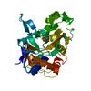
Type: D-peptide linking / Mass: 105.093 Da / Num. of mol.: 1 / Source method: obtained synthetically / Formula: C3H7NO3
#3: Water ChemComp-HOH / water / Water


Mass: 18.015 Da / Num. of mol.: 353 / Source method: isolated from a natural source / Formula: H2O

-
Experimental details

-
Experiment

ExperimentMethod: X-RAY DIFFRACTION / Number of used crystals: 1

-
Sample preparation

CrystalDensity Matthews: 2.2 Å3/Da / Density % sol: 44.11 %
Crystal growTemperature: 277 K / Method: vapor diffusion, hanging drop / pH: 6
Details: PEG 1000, sodium cacodylate, lithium sulfate, pH 6.0, VAPOR DIFFUSION, HANGING DROP, temperature 277K
Crystal grow
*PLUS
Temperature: 4 ℃ / Method: vapor diffusion, hanging drop
Components of the solutions
*PLUS
IDConc.Common nameCrystal-IDSol-IDDetailsChemical formula
1100 mMsodium cacodylate1reservoirpH6.0
2100 mM1reservoirLi2SO4
320 %PEG10001reservoir

-
Data collection

DiffractionMean temperature: 110 K
Diffraction sourceSource: SYNCHROTRON / Site: NSLS / Beamline: X4A / Wavelength: 0.9795 Å
DetectorType: ADSC QUANTUM 4 / Detector: CCD / Date: Aug 9, 2002
RadiationProtocol: SINGLE WAVELENGTH / Monochromatic (M) / Laue (L): M / Scattering type: x-ray
Radiation wavelengthWavelength: 0.9795 Å / Relative weight: 1
ReflectionResolution: 1.45→20 Å / Num. all: 52896 / Num. obs: 51309 / % possible obs: 97 % / Observed criterion σ(I): 14.06 / Biso Wilson estimate: 16.3 Å2
Reflection shellResolution: 1.45→1.5 Å / % possible all: 85.2
Reflection
*PLUS
Num. measured all: 414093 / Rmerge(I) obs: 0.066
Reflection shell
*PLUS
% possible obs: 85.2 % / Rmerge(I) obs: 0.172

-
Processing

Software
NameClassification
DENZOdata reduction
SCALEPACKdata scaling
CNSrefinement
CNSphasing
RefinementMethod to determine structure: FOURIER SYNTHESIS / Resolution: 1.45→9.99 Å / Rfactor Rfree error: 0.003 / Isotropic thermal model: RESTRAINED / Cross valid method: THROUGHOUT / σ(F): 0
RfactorNum. reflection% reflectionSelection details
Rfree0.226 5105 10.1 %RANDOM
Rwork0.204 ---
all0.2041 51236 --
obs0.2041 50626 95.9 %-
Solvent computationSolvent model: FLAT MODEL / Bsol: 56.2052 Å2 / ksol: 0.465542 e/Å3
Displacement parametersBiso mean: 17.8 Å2
Refine analyzeLuzzati coordinate error free: 0.19 Å / Luzzati sigma a free: 0.08 Å
Refinement stepCycle: LAST / Resolution: 1.45→9.99 Å
ProteinNucleic acidLigandSolventTotal
Num. atoms2171 0 7 353 2531
Refine LS restraints
Refine-IDTypeDev idealDev ideal target
X-RAY DIFFRACTIONc_bond_d0.005
X-RAY DIFFRACTIONc_bond_d_na
X-RAY DIFFRACTIONc_bond_d_prot
X-RAY DIFFRACTIONc_angle_d
X-RAY DIFFRACTIONc_angle_d_na
X-RAY DIFFRACTIONc_angle_d_prot
X-RAY DIFFRACTIONc_angle_deg1.2
X-RAY DIFFRACTIONc_angle_deg_na
X-RAY DIFFRACTIONc_angle_deg_prot
X-RAY DIFFRACTIONc_dihedral_angle_d23.2
X-RAY DIFFRACTIONc_dihedral_angle_d_na
X-RAY DIFFRACTIONc_dihedral_angle_d_prot
X-RAY DIFFRACTIONc_improper_angle_d0.78
X-RAY DIFFRACTIONc_improper_angle_d_na
X-RAY DIFFRACTIONc_improper_angle_d_prot
X-RAY DIFFRACTIONc_mcbond_it1.061.5
X-RAY DIFFRACTIONc_mcangle_it1.652
X-RAY DIFFRACTIONc_scbond_it1.952
X-RAY DIFFRACTIONc_scangle_it2.712.5
LS refinement shellResolution: 1.45→1.54 Å / Rfactor Rfree error: 0.009 / Total num. of bins used: 6
RfactorNum. reflection% reflection
Rfree0.234 740 10.1 %
Rwork0.218 6571 -
obs-6571 84.3 %
Xplor file
Refine-IDSerial noParam fileTopol file
X-RAY DIFFRACTION1PROTEIN_REP.PARAMPROTEIN.TOP
X-RAY DIFFRACTION2DS.PARAMDS.TOP
X-RAY DIFFRACTION3WATER_REP.PARAMWATER.TOP
Refinement
*PLUS
Lowest resolution: 10 Å
Solvent computation
*PLUS
Displacement parameters
*PLUS
Refine LS restraints
*PLUS
Refine-IDTypeDev ideal
X-RAY DIFFRACTIONc_angle_deg1.17
X-RAY DIFFRACTIONc_dihedral_angle_d
X-RAY DIFFRACTIONc_dihedral_angle_deg23.2
X-RAY DIFFRACTIONc_improper_angle_d
X-RAY DIFFRACTIONc_improper_angle_deg0.78

+
About Yorodumi

-
News

-
Feb 9, 2022. New format data for meta-information of EMDB entries

New format data for meta-information of EMDB entries

  • Version 3 of the EMDB header file is now the official format.
  • The previous official version 1.9 will be removed from the archive.

Related info.:EMDB header

External links:wwPDB to switch to version 3 of the EMDB data model

-
Aug 12, 2020. Covid-19 info

Covid-19 info

URL: https://pdbj.org/emnavi/covid19.php

New page: Covid-19 featured information page in EM Navigator.

Related info.:Covid-19 info / Mar 5, 2020. Novel coronavirus structure data

+
Mar 5, 2020. Novel coronavirus structure data

Novel coronavirus structure data

Related info.:Yorodumi Speices / Aug 12, 2020. Covid-19 info

External links:COVID-19 featured content - PDBj / Molecule of the Month (242):Coronavirus Proteases

+
Jan 31, 2019. EMDB accession codes are about to change! (news from PDBe EMDB page)

EMDB accession codes are about to change! (news from PDBe EMDB page)

  • The allocation of 4 digits for EMDB accession codes will soon come to an end. Whilst these codes will remain in use, new EMDB accession codes will include an additional digit and will expand incrementally as the available range of codes is exhausted. The current 4-digit format prefixed with “EMD-” (i.e. EMD-XXXX) will advance to a 5-digit format (i.e. EMD-XXXXX), and so on. It is currently estimated that the 4-digit codes will be depleted around Spring 2019, at which point the 5-digit format will come into force.
  • The EM Navigator/Yorodumi systems omit the EMD- prefix.

Related info.:Q: What is EMD? / ID/Accession-code notation in Yorodumi/EM Navigator

External links:EMDB Accession Codes are Changing Soon! / Contact to PDBj

+
Jul 12, 2017. Major update of PDB

Major update of PDB

  • wwPDB released updated PDB data conforming to the new PDBx/mmCIF dictionary.
  • This is a major update changing the version number from 4 to 5, and with Remediation, in which all the entries are updated.
  • In this update, many items about electron microscopy experimental information are reorganized (e.g. em_software).
  • Now, EM Navigator and Yorodumi are based on the updated data.

External links:wwPDB Remediation / Enriched Model Files Conforming to OneDep Data Standards Now Available in the PDB FTP Archive

-
Yorodumi

Thousand views of thousand structures

  • Yorodumi is a browser for structure data from EMDB, PDB, SASBDB, etc.
  • This page is also the successor to EM Navigator detail page, and also detail information page/front-end page for Omokage search.
  • The word "yorodu" (or yorozu) is an old Japanese word meaning "ten thousand". "mi" (miru) is to see.

Related info.:EMDB / PDB / SASBDB / Comparison of 3 databanks / Yorodumi Search / Aug 31, 2016. New EM Navigator & Yorodumi / Yorodumi Papers / Jmol/JSmol / Function and homology information / Changes in new EM Navigator and Yorodumi

Read more