 Movie
Movie Controller

[English] 日本語
 Yorodumi
- PDB-1p91: Crystal Structure of RlmA(I) enzyme: 23S rRNA n1-G745 methyltrans... -
+ Open data

- Basic information
| Entry | Database: PDB / ID: 1p91 | ||||||
|---|---|---|---|---|---|---|---|
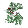
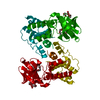
| Title | Crystal Structure of RlmA(I) enzyme: 23S rRNA n1-G745 methyltransferase (NORTHEAST STRUCTURAL GENOMICS CONSORTIUM TARGET ER19) | ||||||
 Components Components | Ribosomal RNA large subunit methyltransferase A | ||||||
 Keywords Keywords | TRANSFERASE / RlmA / RrmA / methyltransferase / G745 / ER19 / 23S rRNA / NESG / STRUCTURAL GENOMICS / PSI / Protein Structure Initiative / Northeast Structural Genomics Consortium | ||||||
| Function / homology |  Function and homology information23S rRNA (guanine745-N1)-methyltransferase / 23S rRNA (guanine(745)-N(1))-methyltransferase activity / rRNA (guanine-N1-)-methyltransferase activity / rRNA base methylation / methyltransferase activity / zinc ion binding Function and homology information23S rRNA (guanine745-N1)-methyltransferase / 23S rRNA (guanine(745)-N(1))-methyltransferase activity / rRNA (guanine-N1-)-methyltransferase activity / rRNA base methylation / methyltransferase activity / zinc ion bindingSimilarity search - Function | ||||||
| Biological species |  Escherichia coli (E. coli) Escherichia coli (E. coli) | ||||||
| Method | X-RAY DIFFRACTION / SYNCHROTRON / MAD / Resolution: 2.8 Å | ||||||
 Authors Authors | Das, K. / Acton, T. / Montelione, G. / Arnold, E. / Northeast Structural Genomics Consortium (NESG) | ||||||
 Citation Citation | Journal: Proc.Natl.Acad.Sci.Usa / Year: 2004 Title: Crystal structure of RlmAI: implications for understanding the 23S rRNA G745/G748-methylation at the macrolide antibiotic-binding site. Authors: Das, K. / Acton, T. / Chiang, Y. / Shih, L. / Arnold, E. / Montelione, G.T. | ||||||
| History |
|
- Structure visualization
| Structure viewer | Molecule: MolmilJmol/JSmol |
|---|
- Downloads & links
-Download
| PDBx/mmCIF format | 1p91.cif.gz | 116.1 KB | Display | PDBx/mmCIF format |
|---|---|---|---|---|
| PDB format | pdb1p91.ent.gz | 96.4 KB | Display | PDB format |
| PDBx/mmJSON format | 1p91.json.gz | Tree view | PDBx/mmJSON format | |
| Others |  Other downloads Other downloads |
-Validation report
| Arichive directory | https://data.pdbj.org/pub/pdb/validation_reports/p9/1p91ftp://data.pdbj.org/pub/pdb/validation_reports/p9/1p91 | HTTPS FTP |
|---|
-Related structure data
| Similar structure data | |
|---|---|
| Other databases |
-Links
 PDBj
PDBj
- Assembly
| Deposited unit | 
| ||||||||
|---|---|---|---|---|---|---|---|---|---|
| 1 |
| ||||||||
| Unit cell |
| ||||||||
| Details | The biological assembly is a dimer. One monomers is related to the other by ~159 degree rotation about the NCS axis. |
-Components
| #1: Protein | / rRNA (guanine-N(1)-)-methyltransferase / 23S rRNA n1-G745 methyltransferase Mass: 30788.188 Da / Num. of mol.: 2 Source method: isolated from a genetically manipulated source Source: (gene. exp.) Escherichia coli (E. coli) / Gene: RRMA OR B1822 / Production host: Escherichia coli (E. coli) / References: UniProt: P36999, EC: 2.1.1.51#2: Chemical |   Mass: 65.409 Da / Num. of mol.: 2 / Source method: obtained synthetically / Formula: Zn Mass: 65.409 Da / Num. of mol.: 2 / Source method: obtained synthetically / Formula: Zn#3: Chemical | ChemComp-SO4 / Sulfate  Mass: 96.063 Da / Num. of mol.: 5 / Source method: obtained synthetically / Formula: SO4 Mass: 96.063 Da / Num. of mol.: 5 / Source method: obtained synthetically / Formula: SO4#4: Chemical | S-Adenosyl methionine  Mass: 398.437 Da / Num. of mol.: 2 / Source method: obtained synthetically / Formula: C15H22N6O5S Mass: 398.437 Da / Num. of mol.: 2 / Source method: obtained synthetically / Formula: C15H22N6O5S#5: Water | ChemComp-HOH / | Water Mass: 18.015 Da / Num. of mol.: 85 / Source method: isolated from a natural source / Formula: H2O Mass: 18.015 Da / Num. of mol.: 85 / Source method: isolated from a natural source / Formula: H2O |
|---|
-Experimental details
-Experiment
| Experiment | Method: X-RAY DIFFRACTION / Number of used crystals: 2 |
|---|
- Sample preparation
| Crystal | Density Matthews: 3.81 Å3/Da / Density % sol: 67.68 % | ||||||||||||||||||||||||||||||||||||||||||||||||
|---|---|---|---|---|---|---|---|---|---|---|---|---|---|---|---|---|---|---|---|---|---|---|---|---|---|---|---|---|---|---|---|---|---|---|---|---|---|---|---|---|---|---|---|---|---|---|---|---|---|
| Crystal grow | Temperature: 300 K / Method: vapor diffusion / pH: 6.5 Details: Ammonium sulfate, Lithium Sulfate, pH 6.5, VAPOR DIFFUSION, temperature 300K | ||||||||||||||||||||||||||||||||||||||||||||||||
| Crystal grow | *PLUS pH: 7.5 / Method: vapor diffusion, hanging drop | ||||||||||||||||||||||||||||||||||||||||||||||||
| Components of the solutions | *PLUS
|
-Data collection
| Diffraction |
| |||||||||||||||
|---|---|---|---|---|---|---|---|---|---|---|---|---|---|---|---|---|
| Diffraction source |
| |||||||||||||||
| Detector |
| |||||||||||||||
| Radiation | Protocol: MAD / Monochromatic (M) / Laue (L): M / Scattering type: x-ray | |||||||||||||||
| Radiation wavelength |
| |||||||||||||||
| Reflection | Resolution: 2.8→50 Å / Num. all: 21876 / Num. obs: 21876 / % possible obs: 93 % / Observed criterion σ(I): -0.5 / Biso Wilson estimate: 41.8 Å2 / Rmerge(I) obs: 0.106 / Net I/σ(I): 11 | |||||||||||||||
| Reflection shell | Resolution: 2.8→2.85 Å / % possible all: 80.2 | |||||||||||||||
| Reflection | *PLUS Lowest resolution: 50 Å / % possible obs: 93 % / Num. measured all: 88268 |

- Processing
| Software |
| |||||||||||||||||||||||||
|---|---|---|---|---|---|---|---|---|---|---|---|---|---|---|---|---|---|---|---|---|---|---|---|---|---|---|
| Refinement | Method to determine structure: MAD / Resolution: 2.8→20 Å / Rfactor Rfree error: 0.009 / Data cutoff high absF: 2974439.24 / Data cutoff high rms absF: 2974439.24 / Data cutoff low absF: 0 / Isotropic thermal model: RESTRAINED / Cross valid method: THROUGHOUT / σ(F): 1 / Stereochemistry target values: Engh & Huber
| |||||||||||||||||||||||||
| Solvent computation | Solvent model: FLAT MODEL / Bsol: 10 Å2 / ksol: 0.251692 e/Å3 | |||||||||||||||||||||||||
| Displacement parameters | Biso mean: 55.8 Å2
| |||||||||||||||||||||||||
| Refine analyze |
| |||||||||||||||||||||||||
| Refinement step | Cycle: LAST / Resolution: 2.8→20 Å
| |||||||||||||||||||||||||
| Refine LS restraints |
| |||||||||||||||||||||||||
| LS refinement shell | Resolution: 2.8→2.97 Å / Rfactor Rfree error: 0.035 / Total num. of bins used: 6
| |||||||||||||||||||||||||
| Xplor file |
| |||||||||||||||||||||||||
| Refinement | *PLUS Highest resolution: 2.8 Å / Lowest resolution: 20 Å / % reflection Rfree: 5 % | |||||||||||||||||||||||||
| Solvent computation | *PLUS | |||||||||||||||||||||||||
| Displacement parameters | *PLUS | |||||||||||||||||||||||||
| Refine LS restraints | *PLUS
|
