 Movie
Movie Controller

[English] 日本語
 Yorodumi
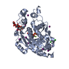
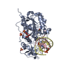
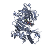
- PDB-1p4n: Crystal Structure of Weissella viridescens FemX:UDP-MurNAc-pentap... -
+ Open data

- Basic information
| Entry | Database: PDB / ID: 1p4n | ||||||
|---|---|---|---|---|---|---|---|
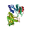
| Title | Crystal Structure of Weissella viridescens FemX:UDP-MurNAc-pentapeptide complex | ||||||
 Components Components |
| ||||||
 Keywords Keywords | Transferase/Transferase Substrate /  FemX / Transferase-Transferase Substrate complex FemX / Transferase-Transferase Substrate complex | ||||||
| Function / homology |  Function and homology informationUDP-N-acetylmuramoylpentapeptide-lysine N6-alanyltransferase / UDP-N-acetylmuramoylpentapeptide-lysine N6-alanyltransferase activity / peptidoglycan biosynthetic process / cell wall organization / regulation of cell shape Function and homology informationUDP-N-acetylmuramoylpentapeptide-lysine N6-alanyltransferase / UDP-N-acetylmuramoylpentapeptide-lysine N6-alanyltransferase activity / peptidoglycan biosynthetic process / cell wall organization / regulation of cell shapeSimilarity search - Function | ||||||
| Biological species |  Weissella viridescens (bacteria) Weissella viridescens (bacteria) Staphylococcus aureus (bacteria) Staphylococcus aureus (bacteria) | ||||||
| Method | X-RAY DIFFRACTION / SYNCHROTRON / difference fourier synthesis / Resolution: 1.9 Å | ||||||
 Authors Authors | Biarrotte-Sorin, S. / Maillard, A. / Delettre, J. / Sougakoff, W. / Arthur, M. / Mayer, C. | ||||||
 Citation Citation | Journal: Structure / Year: 2004 Title: Crystal structures of Weissella viridescens FemX and its complex with UDP-MurNAc-pentapeptide: insights into FemABX family substrates recognition. Authors: Biarrotte-Sorin, S. / Maillard, A.P. / Delettre, J. / Sougakoff, W. / Arthur, M. / Mayer, C. | ||||||
| History |
| ||||||
| Remark 600 | HETEROGEN The residue FGA is gamma-glutamic acid but does not make classical peptide bond. It makes ...HETEROGEN The residue FGA is gamma-glutamic acid but does not make classical peptide bond. It makes peptide bond on the side chain at the Cgamma. |
- Structure visualization
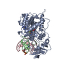
| Structure viewer | Molecule: MolmilJmol/JSmol |
|---|
- Downloads & links
-Download
| PDBx/mmCIF format | 1p4n.cif.gz | 96.3 KB | Display | PDBx/mmCIF format |
|---|---|---|---|---|
| PDB format | pdb1p4n.ent.gz | 70 KB | Display | PDB format |
| PDBx/mmJSON format | 1p4n.json.gz | Tree view | PDBx/mmJSON format | |
| Others |  Other downloads Other downloads |
-Validation report
| Arichive directory | https://data.pdbj.org/pub/pdb/validation_reports/p4/1p4nftp://data.pdbj.org/pub/pdb/validation_reports/p4/1p4n | HTTPS FTP |
|---|
-Related structure data
| Related structure data |  1ne9SC S: Starting model for refinement C: citing same article ( |
|---|---|
| Similar structure data |


-Links
 PDBj
PDBj

- Assembly
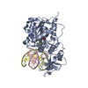
| Deposited unit | 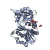
| ||||||||
|---|---|---|---|---|---|---|---|---|---|
| 1 |
| ||||||||
| Unit cell |
|
-Components
| #1: Protein | Lipid II:glycine glycyltransferase Mass: 38195.801 Da / Num. of mol.: 1 Source method: isolated from a genetically manipulated source Source: (gene. exp.) Weissella viridescens (bacteria) / Gene: femx / Plasmid: pTYB2 / Production host: Escherichia coli (E. coli) / Strain (production host): ER2566References: GenBank: 11999275, UniProt: Q9EY50*PLUS, UDP-N-acetylmuramoylpentapeptide-lysine N6-alanyltransferase | ||||
|---|---|---|---|---|---|
| #2: Protein/peptide | Mass: 1150.943 Da / Num. of mol.: 1 / Source method: isolated from a natural source / Source: (natural) Staphylococcus aureus (bacteria) | ||||
| #3: Chemical |   Mass: 24.305 Da / Num. of mol.: 3 / Source method: obtained synthetically / Formula: Mg Mass: 24.305 Da / Num. of mol.: 3 / Source method: obtained synthetically / Formula: Mg#4: Chemical | Glycerol  Mass: 92.094 Da / Num. of mol.: 2 / Source method: obtained synthetically / Formula: C3H8O3 Mass: 92.094 Da / Num. of mol.: 2 / Source method: obtained synthetically / Formula: C3H8O3#5: Water | ChemComp-HOH / | Water Mass: 18.015 Da / Num. of mol.: 466 / Source method: isolated from a natural source / Formula: H2O Mass: 18.015 Da / Num. of mol.: 466 / Source method: isolated from a natural source / Formula: H2O |
-Experimental details
-Experiment
| Experiment | Method: X-RAY DIFFRACTION / Number of used crystals: 1 |
|---|
- Sample preparation
| Crystal | Density Matthews: 2.23 Å3/Da / Density % sol: 44.8 % | |||||||||||||||||||||||||||||||||||
|---|---|---|---|---|---|---|---|---|---|---|---|---|---|---|---|---|---|---|---|---|---|---|---|---|---|---|---|---|---|---|---|---|---|---|---|---|
| Crystal grow | Temperature: 298 K / Method: vapor diffusion, hanging drop / pH: 6.5 Details: PEG 2K, NaCl, sodium cacodylate, pH 6.5, VAPOR DIFFUSION, HANGING DROP, temperature 298K | |||||||||||||||||||||||||||||||||||
| Crystal grow | *PLUS Method: vapor diffusion | |||||||||||||||||||||||||||||||||||
| Components of the solutions | *PLUS
|
-Data collection
| Diffraction | Mean temperature: 100 K |
|---|---|
| Diffraction source | Source: SYNCHROTRON / Site: ESRF  / Beamline: BM30A / Wavelength: 0.979711 Å / Beamline: BM30A / Wavelength: 0.979711 Å |
| Detector | Type: MARRESEARCH / Detector: CCD / Date: Dec 14, 2002 Details: two crystals monochromator between two cylindrical parabolic mirrors |
| Radiation | Monochromator: Sagitally focused Si (111) / Protocol: SINGLE WAVELENGTH / Monochromatic (M) / Laue (L): M / Scattering type: x-ray |
| Radiation wavelength | Wavelength: 0.979711 Å / Relative weight: 1 |
| Reflection | Resolution: 1.9→15.57 Å / Num. obs: 27221 / % possible obs: 98.6 % / Observed criterion σ(F): 0 / Observed criterion σ(I): 0 / Redundancy: 3.6 % / Biso Wilson estimate: 12.7 Å2 / Rsym value: 0.073 / Net I/σ(I): 7.5 |
| Reflection shell | Resolution: 1.9→2 Å / Redundancy: 3.8 % / Mean I/σ(I) obs: 3.7 / Num. unique all: 3947 / Rsym value: 0.19 / % possible all: 98 |
| Reflection | *PLUS Num. measured all: 102033 / Rmerge(I) obs: 0.073 |
| Reflection shell | *PLUS % possible obs: 98 % / Rmerge(I) obs: 0.19 |
- Processing
| Software |
| |||||||||||||||||||||||||||
|---|---|---|---|---|---|---|---|---|---|---|---|---|---|---|---|---|---|---|---|---|---|---|---|---|---|---|---|---|
| Refinement | Method to determine structure: difference fourier synthesis Starting model: PDB ENTRY 1NE9 Resolution: 1.9→15.57 Å / Rfactor Rfree error: 0.006 / Data cutoff high absF: 1090877 / Data cutoff high rms absF: 1090877 / Data cutoff low absF: 0 / Isotropic thermal model: OVERALL / Cross valid method: THROUGHOUT / σ(F): 0 / σ(I): 0 / Stereochemistry target values: Engh & Huber
| |||||||||||||||||||||||||||
| Solvent computation | Solvent model: FLAT MODEL / Bsol: 54.1592 Å2 / ksol: 0.39732 e/Å3 | |||||||||||||||||||||||||||
| Displacement parameters | Biso mean: 16.3 Å2
| |||||||||||||||||||||||||||
| Refine analyze |
| |||||||||||||||||||||||||||
| Refinement step | Cycle: LAST / Resolution: 1.9→15.57 Å
| |||||||||||||||||||||||||||
| Refine LS restraints |
| |||||||||||||||||||||||||||
| LS refinement shell | Resolution: 1.9→2 Å / Rfactor Rfree error: 0.018 / Total num. of bins used: 6
| |||||||||||||||||||||||||||
| Xplor file |
| |||||||||||||||||||||||||||
| Refinement | *PLUS Highest resolution: 1.9 Å | |||||||||||||||||||||||||||
| Solvent computation | *PLUS | |||||||||||||||||||||||||||
| Displacement parameters | *PLUS | |||||||||||||||||||||||||||
| Refine LS restraints | *PLUS
| |||||||||||||||||||||||||||
| LS refinement shell | *PLUS Highest resolution: 1.9 Å / Lowest resolution: 2 Å |
