 Movie
Movie Controller

[English] 日本語
 Yorodumi
- PDB-1ozg: The crystal structure of Klebsiella pneumoniae acetolactate synth... -
+ Open data

- Basic information
| Entry | Database: PDB / ID: 1ozg | ||||||
|---|---|---|---|---|---|---|---|
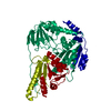
| Title | The crystal structure of Klebsiella pneumoniae acetolactate synthase with enzyme-bound cofactor and with an unusual intermediate | ||||||
 Components Components | Acetolactate synthase, catabolic | ||||||
 Keywords Keywords | LYASE / acetolactate synthase / acetohydroxyacid synthase / thiamin diphosphate | ||||||
| Function / homology |  Function and homology information Function and homology informationbutanediol metabolic process / acetolactate synthase / acetolactate synthase activity / carboxylic acid metabolic process / thiamine pyrophosphate binding / magnesium ion bindingSimilarity search - Function | ||||||
| Biological species |  Klebsiella pneumoniae (bacteria) Klebsiella pneumoniae (bacteria) | ||||||
| Method | X-RAY DIFFRACTION / SYNCHROTRON / MOLECULAR REPLACEMENT / Resolution: 2.3 Å | ||||||
 Authors Authors | Pang, S.S. / Duggleby, R.G. / Schowen, R.L. / Guddat, L.W. | ||||||
 Citation Citation | Journal: J.Biol.Chem. / Year: 2004 Title: The Crystal Structures of Klebsiella pneumoniae Acetolactate Synthase with Enzyme-bound Cofactor and with an Unusual Intermediate. Authors: Pang, S.S. / Duggleby, R.G. / Schowen, R.L. / Guddat, L.W. | ||||||
| History |
|
- Structure visualization
| Structure viewer | Molecule: MolmilJmol/JSmol |
|---|
- Downloads & links
-Download
| PDBx/mmCIF format | 1ozg.cif.gz | 235.1 KB | Display | PDBx/mmCIF format |
|---|---|---|---|---|
| PDB format | pdb1ozg.ent.gz | 185.6 KB | Display | PDB format |
| PDBx/mmJSON format | 1ozg.json.gz | Tree view | PDBx/mmJSON format | |
| Others |  Other downloads Other downloads |
-Validation report
| Arichive directory | https://data.pdbj.org/pub/pdb/validation_reports/oz/1ozgftp://data.pdbj.org/pub/pdb/validation_reports/oz/1ozg | HTTPS FTP |
|---|
-Related structure data
| Related structure data |  1n0hC 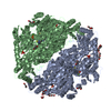 1ozfC 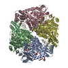 1ozhC  1jscS C: citing same article ( S: Starting model for refinement |
|---|---|
| Similar structure data |


-Links
 PDBj
PDBj


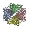
- Assembly
| Deposited unit | 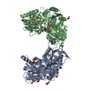
| ||||||||
|---|---|---|---|---|---|---|---|---|---|
| 1 | 
| ||||||||
| Unit cell |
| ||||||||
| Details | The biological assembly is a tetramer generated from the dimer in the asymmetric unit by the operation: -X,Y,-Z+1/2 |
-Components
-Protein , 1 types, 2 molecules AB
| #1: Protein | / ALS Mass: 61355.766 Da / Num. of mol.: 2 Source method: isolated from a genetically manipulated source Source: (gene. exp.) Klebsiella pneumoniae (bacteria) / Gene: BUDB OR ILVK / Plasmid: pET30a(+) / Species (production host): Escherichia coli / Production host: Escherichia coli BL21(DE3) (bacteria) / Strain (production host): BL21(DE3) / References: UniProt: P27696, EC: 4.1.3.18 |
|---|
-Non-polymers , 5 types, 626 molecules 


| #2: Chemical | Phosphate Mass: 94.971 Da / Num. of mol.: 2 / Source method: obtained synthetically / Formula: PO4 Mass: 94.971 Da / Num. of mol.: 2 / Source method: obtained synthetically / Formula: PO4#3: Chemical |  Mass: 24.305 Da / Num. of mol.: 2 / Source method: obtained synthetically / Formula: Mg Mass: 24.305 Da / Num. of mol.: 2 / Source method: obtained synthetically / Formula: Mg#4: Chemical |  Mass: 468.359 Da / Num. of mol.: 2 / Source method: obtained synthetically / Formula: C14H22N4O8P2S Mass: 468.359 Da / Num. of mol.: 2 / Source method: obtained synthetically / Formula: C14H22N4O8P2S#5: Chemical | ChemComp-PEG / Diethylene glycol Mass: 106.120 Da / Num. of mol.: 8 / Source method: obtained synthetically / Formula: C4H10O3 Mass: 106.120 Da / Num. of mol.: 8 / Source method: obtained synthetically / Formula: C4H10O3#6: Water | ChemComp-HOH / | WaterMass: 18.015 Da / Num. of mol.: 612 / Source method: isolated from a natural source / Formula: H2O |
|---|
-Experimental details
-Experiment
| Experiment | Method: X-RAY DIFFRACTION / Number of used crystals: 1 |
|---|
- Sample preparation
| Crystal | Density Matthews: 2.42 Å3/Da / Density % sol: 48.86 % | |||||||||||||||||||||||||||||||||||||||||||||||||||||||||||||||
|---|---|---|---|---|---|---|---|---|---|---|---|---|---|---|---|---|---|---|---|---|---|---|---|---|---|---|---|---|---|---|---|---|---|---|---|---|---|---|---|---|---|---|---|---|---|---|---|---|---|---|---|---|---|---|---|---|---|---|---|---|---|---|---|---|
| Crystal grow | Temperature: 290 K / Method: vapor diffusion, hanging drop / pH: 7.6 Details: PEG 8000, ethylene glycol, sodium HEPES , pH 7.6, VAPOR DIFFUSION, HANGING DROP, temperature 290K | |||||||||||||||||||||||||||||||||||||||||||||||||||||||||||||||
| Crystal grow | *PLUS Temperature: 17 ℃ / pH: 7 / Method: vapor diffusion, hanging drop | |||||||||||||||||||||||||||||||||||||||||||||||||||||||||||||||
| Components of the solutions | *PLUS
|
-Data collection
| Diffraction | Mean temperature: 100 K |
|---|---|
| Diffraction source | Source: SYNCHROTRON / Site: APS  / Beamline: 14-BM-D / Wavelength: 1 Å / Beamline: 14-BM-D / Wavelength: 1 Å |
| Detector | Type: ADSC QUANTUM 4 / Detector: CCD / Date: Feb 22, 2002 / Details: mirrors |
| Radiation | Monochromator: Si 111 / Protocol: SINGLE WAVELENGTH / Monochromatic (M) / Laue (L): M / Scattering type: x-ray |
| Radiation wavelength | Wavelength: 1 Å / Relative weight: 1 |
| Reflection | Resolution: 2.3→100 Å / Num. all: 48466 / Num. obs: 48466 / % possible obs: 88.2 % / Observed criterion σ(F): 0 / Observed criterion σ(I): 0 / Redundancy: 3.5 % / Rsym value: 0.057 / Net I/σ(I): 17.7 |
| Reflection shell | Resolution: 2.3→2.38 Å / Redundancy: 1.7 % / Mean I/σ(I) obs: 4.5 / Num. unique all: 3030 / Rsym value: 0.126 / % possible all: 49.3 |
| Reflection | *PLUS Lowest resolution: 100 Å / Num. measured all: 170273 / Rmerge(I) obs: 0.057 |
| Reflection shell | *PLUS % possible obs: 49.3 % / Num. unique obs: 3030 / Num. measured obs: 5047 / Rmerge(I) obs: 0.126 |
- Processing
| Software |
| |||||||||||||||||||||||||
|---|---|---|---|---|---|---|---|---|---|---|---|---|---|---|---|---|---|---|---|---|---|---|---|---|---|---|
| Refinement | Method to determine structure: MOLECULAR REPLACEMENT Starting model: PDB entry 1JSC Resolution: 2.3→100 Å / Isotropic thermal model: anisotropic / Cross valid method: THROUGHOUT / σ(F): 0 / σ(I): 0 / Stereochemistry target values: Engh & Huber
| |||||||||||||||||||||||||
| Displacement parameters | Biso mean: 26.92 Å2
| |||||||||||||||||||||||||
| Refine analyze |
| |||||||||||||||||||||||||
| Refinement step | Cycle: LAST / Resolution: 2.3→100 Å
| |||||||||||||||||||||||||
| Refine LS restraints |
| |||||||||||||||||||||||||
| LS refinement shell | Resolution: 2.3→2.38 Å
| |||||||||||||||||||||||||
| Refinement | *PLUS Lowest resolution: 100 Å | |||||||||||||||||||||||||
| Solvent computation | *PLUS | |||||||||||||||||||||||||
| Displacement parameters | *PLUS | |||||||||||||||||||||||||
| Refine LS restraints | *PLUS
|
