 Movie
Movie Controller

[English] 日本語
 Yorodumi
- PDB-1oz9: Crystal structure of AQ_1354, a hypothetical protein from Aquifex... -
+ Open data

- Basic information
| Entry | Database: PDB / ID: 1oz9 | ||||||
|---|---|---|---|---|---|---|---|
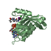
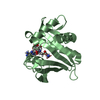
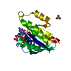
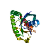
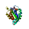
| Title | Crystal structure of AQ_1354, a hypothetical protein from Aquifex aeolicus | ||||||
 Components Components | Hypothetical protein AQ_1354 Hypothesis Hypothesis | ||||||
 Keywords Keywords | UNKNOWN FUNCTION / matrix metalloproteinase type fold / Structural Genomics / BSGC structure funded by NIH / Protein Structure Initiative / PSI / Berkeley Structural Genomics Center | ||||||
| Function / homology |  Function and homology information Function and homology informationRNA endonuclease activity / metalloendopeptidase activity / rRNA processing / Hydrolases; Acting on ester bonds / zinc ion binding / cytoplasmSimilarity search - Function | ||||||
| Biological species |   Aquifex aeolicus (bacteria) Aquifex aeolicus (bacteria) | ||||||
| Method | X-RAY DIFFRACTION / SYNCHROTRON / SAD / Resolution: 1.894 Å | ||||||
 Authors Authors | Oganesyan, V. / Busso, D. / Brandsen, J. / Chen, S. / Jancarik, J. / Kim, R. / Kim, S.H. / Berkeley Structural Genomics Center (BSGC) | ||||||
 Citation Citation | Journal: Acta Crystallogr.,Sect.D / Year: 2003 Title: Structure of the hypothetical protein AQ_1354 from Aquifex aeolicus. Authors: Oganesyan, V. / Busso, D. / Brandsen, J. / Chen, S. / Jancarik, J. / Kim, R. / Kim, S.H. | ||||||
| History |
|
- Structure visualization
| Structure viewer | Molecule: MolmilJmol/JSmol |
|---|
- Downloads & links
-Download
| PDBx/mmCIF format | 1oz9.cif.gz | 40.7 KB | Display | PDBx/mmCIF format |
|---|---|---|---|---|
| PDB format | pdb1oz9.ent.gz | 29.4 KB | Display | PDB format |
| PDBx/mmJSON format | 1oz9.json.gz | Tree view | PDBx/mmJSON format | |
| Others |  Other downloads Other downloads |
-Validation report
| Arichive directory | https://data.pdbj.org/pub/pdb/validation_reports/oz/1oz9ftp://data.pdbj.org/pub/pdb/validation_reports/oz/1oz9 | HTTPS FTP |
|---|
-Related structure data
| Similar structure data | |
|---|---|
| Other databases |


-Links
 PDBj
PDBj- Assembly
| Deposited unit | 
| ||||||||
|---|---|---|---|---|---|---|---|---|---|
| 1 |
| ||||||||
| Unit cell |
| ||||||||
| Details | The biological assembly is a monomer |
-Components
| #1: Protein | Hypothesis Mass: 17372.021 Da / Num. of mol.: 1 Source method: isolated from a genetically manipulated source Source: (gene. exp.) Aquifex aeolicus (bacteria) / Plasmid: pLIC / Species (production host): Escherichia coli / Production host: Escherichia coli BL21 (bacteria) / Strain (production host): BL21 / References: UniProt: O67367 |
|---|---|
| #2: Water | ChemComp-HOH / Water Mass: 18.015 Da / Num. of mol.: 50 / Source method: isolated from a natural source / Formula: H2O Mass: 18.015 Da / Num. of mol.: 50 / Source method: isolated from a natural source / Formula: H2O |
-Experimental details
-Experiment
| Experiment | Method: X-RAY DIFFRACTION / Number of used crystals: 1 |
|---|
- Sample preparation
| Crystal | Density Matthews: 2.76 Å3/Da / Density % sol: 55.07 % | |||||||||||||||||||||||||||||||||||||||||||||||||||||||||||||||
|---|---|---|---|---|---|---|---|---|---|---|---|---|---|---|---|---|---|---|---|---|---|---|---|---|---|---|---|---|---|---|---|---|---|---|---|---|---|---|---|---|---|---|---|---|---|---|---|---|---|---|---|---|---|---|---|---|---|---|---|---|---|---|---|---|
| Crystal grow | Temperature: 298 K / Method: vapor diffusion, hanging drop / pH: 8.5 Details: PEG 4K, Na Acetate, Tris-HCl, pH 8.5, VAPOR DIFFUSION, HANGING DROP, temperature 298.0K | |||||||||||||||||||||||||||||||||||||||||||||||||||||||||||||||
| Crystal grow | *PLUS pH: 7 / Method: vapor diffusion, hanging drop | |||||||||||||||||||||||||||||||||||||||||||||||||||||||||||||||
| Components of the solutions | *PLUS
|
-Data collection
| Diffraction | Mean temperature: 100 K |
|---|---|
| Diffraction source | Source: SYNCHROTRON / Site: ALS  / Beamline: 5.0.2 / Wavelength: 0.9792 Å / Beamline: 5.0.2 / Wavelength: 0.9792 Å |
| Detector | Type: ADSC QUANTUM 4 / Detector: CCD / Date: Aug 14, 2002 / Details: mirrors |
| Radiation | Monochromator: graphite / Protocol: SINGLE WAVELENGTH / Monochromatic (M) / Laue (L): M / Scattering type: x-ray |
| Radiation wavelength | Wavelength: 0.9792 Å / Relative weight: 1 |
| Reflection | Resolution: 1.89→41.17 Å / Num. all: 14413 / Num. obs: 14413 / % possible obs: 100 % / Observed criterion σ(F): 0 / Observed criterion σ(I): 0 / Redundancy: 6.5 % / Biso Wilson estimate: 29 Å2 / Rmerge(I) obs: 0.055 / Net I/σ(I): 25 |
| Reflection shell | Resolution: 1.894→1.943 Å / Redundancy: 5.8 % / Rmerge(I) obs: 0.459 / Mean I/σ(I) obs: 2.2 / Num. unique all: 982 / % possible all: 100 |
| Reflection | *PLUS Lowest resolution: 41.2 Å / Num. obs: 28054 / % possible obs: 96.3 % / Redundancy: 9.3 % |
| Reflection shell | *PLUS Highest resolution: 1.89 Å / Lowest resolution: 1.97 Å / % possible obs: 85.8 % / Rmerge(I) obs: 0.46 / Mean I/σ(I) obs: 2.25 |
- Processing
| Software |
| |||||||||||||||||||||||||||||||||||||||||||||||||||||||||||||||||||||||||||
|---|---|---|---|---|---|---|---|---|---|---|---|---|---|---|---|---|---|---|---|---|---|---|---|---|---|---|---|---|---|---|---|---|---|---|---|---|---|---|---|---|---|---|---|---|---|---|---|---|---|---|---|---|---|---|---|---|---|---|---|---|---|---|---|---|---|---|---|---|---|---|---|---|---|---|---|---|
| Refinement | Method to determine structure: SAD / Resolution: 1.894→12 Å / Cor.coef. Fo:Fc: 0.95 / Cor.coef. Fo:Fc free: 0.937 / SU B: 3.105 / SU ML: 0.091 / TLS residual ADP flag: LIKELY RESIDUAL / Isotropic thermal model: Isotropic / Cross valid method: THROUGHOUT / σ(F): 0 / σ(I): 0 / ESU R: 0.151 / ESU R Free: 0.138 / Stereochemistry target values: MAXIMUM LIKELIHOOD
| |||||||||||||||||||||||||||||||||||||||||||||||||||||||||||||||||||||||||||
| Solvent computation | Ion probe radii: 0.8 Å / Shrinkage radii: 0.8 Å / VDW probe radii: 1.4 Å / Solvent model: BABINET MODEL WITH MASK | |||||||||||||||||||||||||||||||||||||||||||||||||||||||||||||||||||||||||||
| Displacement parameters | Biso mean: 22.066 Å2
| |||||||||||||||||||||||||||||||||||||||||||||||||||||||||||||||||||||||||||
| Refine analyze |
| |||||||||||||||||||||||||||||||||||||||||||||||||||||||||||||||||||||||||||
| Refinement step | Cycle: LAST / Resolution: 1.894→12 Å
| |||||||||||||||||||||||||||||||||||||||||||||||||||||||||||||||||||||||||||
| Refine LS restraints |
| |||||||||||||||||||||||||||||||||||||||||||||||||||||||||||||||||||||||||||
| LS refinement shell | Resolution: 1.894→1.943 Å / Total num. of bins used: 20
| |||||||||||||||||||||||||||||||||||||||||||||||||||||||||||||||||||||||||||
| Refinement TLS params. | Method: refined / Origin x: 47.99 Å / Origin y: 18.021 Å / Origin z: 43.601 Å
| |||||||||||||||||||||||||||||||||||||||||||||||||||||||||||||||||||||||||||
| Software | *PLUS Version: 5 / Classification: refinement | |||||||||||||||||||||||||||||||||||||||||||||||||||||||||||||||||||||||||||
| Refinement | *PLUS Highest resolution: 1.89 Å / Num. reflection obs: 15227 / % reflection Rfree: 5 % / Rfactor Rfree: 0.234 / Rfactor Rwork: 0.201 | |||||||||||||||||||||||||||||||||||||||||||||||||||||||||||||||||||||||||||
| Solvent computation | *PLUS | |||||||||||||||||||||||||||||||||||||||||||||||||||||||||||||||||||||||||||
| Displacement parameters | *PLUS | |||||||||||||||||||||||||||||||||||||||||||||||||||||||||||||||||||||||||||
| Refine LS restraints | *PLUS
|
