[English] 日本語
Yorodumi
- PDB-1oxh: The crystal structure of beta-ketoacyl-[acyl carrier protein] syn... -

+
Open data


ID or keywords:

Loading...

-
Basic information

Entry
Database: PDB / ID: 1oxh
Title
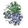
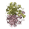
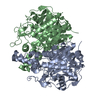
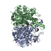
The crystal structure of beta-ketoacyl-[acyl carrier protein] synthase II from Streptococcus Pneumoniae, Triclinic form
ComponentsBeta ketoacyl-acyl carrier protein synthase
KeywordsTRANSFERASE
Function / homology
Function and homology information


beta-ketoacyl-[acyl-carrier-protein] synthase II / 3-oxoacyl-[acyl-carrier-protein] synthase activity / fatty acid biosynthetic process / metal ion binding
Similarity search - Function
3-oxoacyl-[acyl-carrier-protein] synthase 2 / Beta-ketoacyl synthase / Thiolase/Chalcone synthase / Peroxisomal Thiolase; Chain A, domain 1 / Ketosynthase family 3 (KS3) domain profile. / Beta-ketoacyl synthase / Beta-ketoacyl synthase, active site / Ketosynthase family 3 (KS3) active site signature. / Polyketide synthase, beta-ketoacyl synthase domain / Beta-ketoacyl synthase, N-terminal ...3-oxoacyl-[acyl-carrier-protein] synthase 2 / Beta-ketoacyl synthase / Thiolase/Chalcone synthase / Peroxisomal Thiolase; Chain A, domain 1 / Ketosynthase family 3 (KS3) domain profile. / Beta-ketoacyl synthase / Beta-ketoacyl synthase, active site / Ketosynthase family 3 (KS3) active site signature. / Polyketide synthase, beta-ketoacyl synthase domain / Beta-ketoacyl synthase, N-terminal / Beta-ketoacyl synthase, C-terminal / Beta-ketoacyl synthase, N-terminal domain / Beta-ketoacyl synthase, C-terminal domain / Thiolase-like / 3-Layer(aba) Sandwich / Alpha Beta
Similarity search - Domain/homology
3-oxoacyl-[acyl-carrier-protein] synthase 2
Similarity search - Component
Biological speciesStreptococcus pneumoniae (bacteria)
MethodX-RAY DIFFRACTION / SYNCHROTRON / MOLECULAR REPLACEMENT / Resolution: 2.09 Å
AuthorsPrice, A.C. / Rock, C.O. / White, S.W.
CitationJournal: J.Bacteriol. / Year: 2003
Title: The 1.3-Angstrom-Resolution Crystal Structure of beta-Ketoacyl-Acyl Carrier Protein Synthase II from Streptococcus pneumoniae.
Authors: Price, A.C. / Rock, C.O. / White, S.W.
History
DepositionApr 2, 2003Deposition site: RCSB / Processing site: RCSB
Revision 1.0Apr 22, 2003Provider: repository / Type: Initial release
Revision 1.1Apr 29, 2008Group: Version format compliance
Revision 1.2Jul 13, 2011Group: Version format compliance
Revision 1.3Jan 31, 2018Group: Experimental preparation / Category: exptl_crystal_grow / Item: _exptl_crystal_grow.temp
Revision 1.4Feb 14, 2024Group: Data collection / Database references / Derived calculations
Category: chem_comp_atom / chem_comp_bond ...chem_comp_atom / chem_comp_bond / database_2 / pdbx_struct_conn_angle / struct_conn / struct_ref_seq_dif / struct_site
Item: _database_2.pdbx_DOI / _database_2.pdbx_database_accession ..._database_2.pdbx_DOI / _database_2.pdbx_database_accession / _pdbx_struct_conn_angle.ptnr1_auth_comp_id / _pdbx_struct_conn_angle.ptnr1_auth_seq_id / _pdbx_struct_conn_angle.ptnr1_label_atom_id / _pdbx_struct_conn_angle.ptnr1_label_comp_id / _pdbx_struct_conn_angle.ptnr1_label_seq_id / _pdbx_struct_conn_angle.ptnr3_auth_comp_id / _pdbx_struct_conn_angle.ptnr3_auth_seq_id / _pdbx_struct_conn_angle.ptnr3_label_atom_id / _pdbx_struct_conn_angle.ptnr3_label_comp_id / _pdbx_struct_conn_angle.ptnr3_label_seq_id / _pdbx_struct_conn_angle.value / _struct_conn.pdbx_dist_value / _struct_conn.ptnr1_auth_comp_id / _struct_conn.ptnr1_auth_seq_id / _struct_conn.ptnr1_label_asym_id / _struct_conn.ptnr1_label_atom_id / _struct_conn.ptnr1_label_comp_id / _struct_conn.ptnr1_label_seq_id / _struct_conn.ptnr2_auth_comp_id / _struct_conn.ptnr2_auth_seq_id / _struct_conn.ptnr2_label_asym_id / _struct_conn.ptnr2_label_atom_id / _struct_conn.ptnr2_label_comp_id / _struct_conn.ptnr2_label_seq_id / _struct_ref_seq_dif.details / _struct_site.pdbx_auth_asym_id / _struct_site.pdbx_auth_comp_id / _struct_site.pdbx_auth_seq_id
Remark 600HETEROGEN WATERS 102-140 ARE ASSOCIATED WITH CHAIN A. WATERS 202-240 ARE ASSOCIATED WITH CHAIN B. ...HETEROGEN WATERS 102-140 ARE ASSOCIATED WITH CHAIN A. WATERS 202-240 ARE ASSOCIATED WITH CHAIN B. WATERS 302-340 ARE ASSOCIATED WITH CHAIN C. WATERS 402-440 ARE ASSOCIATED WITH CHAIN D.

-
Structure visualization

Structure viewerMolecule:
MolmilJmol/JSmol

Downloads & links

-
Assembly

Deposited unit
A: Beta ketoacyl-acyl carrier protein synthase
B: Beta ketoacyl-acyl carrier protein synthase
C: Beta ketoacyl-acyl carrier protein synthase
D: Beta ketoacyl-acyl carrier protein synthase
hetero molecules


Theoretical massNumber of molelcules
Total (without water)184,1718
Polymers184,0744
Non-polymers974
Water3,891216
1
A: Beta ketoacyl-acyl carrier protein synthase
B: Beta ketoacyl-acyl carrier protein synthase
hetero molecules


Theoretical massNumber of molelcules
Total (without water)92,0864
Polymers92,0372
Non-polymers492
Water362
TypeNameSymmetry operationNumber
identity operation1_555x,y,z1
Buried area5960 Å2
ΔGint-55 kcal/mol
Surface area26050 Å2
MethodPISA
2
C: Beta ketoacyl-acyl carrier protein synthase
D: Beta ketoacyl-acyl carrier protein synthase
hetero molecules


Theoretical massNumber of molelcules
Total (without water)92,0864
Polymers92,0372
Non-polymers492
Water362
TypeNameSymmetry operationNumber
identity operation1_555x,y,z1
Buried area5920 Å2
ΔGint-56 kcal/mol
Surface area26160 Å2
MethodPISA
Unit cell
Length a, b, c (Å)61.520, 71.646, 96.100
Angle α, β, γ (deg.)89.83, 83.09, 69.15
Int Tables number1
Space group name H-MP1
Noncrystallographic symmetry (NCS)NCS domain:
IDEns-IDDetails
11A
21B
31C
41D
51I
61J
71K
81L

NCS domain segments:
Dom-IDComponent-IDEns-IDRefine codeAuth asym-IDAuth seq-ID
1111A3
2111B3
3111C3
4111D3
1211A5 - 10
2211B5 - 10
3211C5 - 10
4211D5 - 10
1311A12
2311B12
3311C12
4311D12
1411A16 - 19
2411B16 - 19
3411C16 - 19
4411D16 - 19
1511A27
2511B27
3511C27
4511D27
1611A34 - 54
2611B34 - 54
3611C34 - 54
4611D34 - 54
1711A57 - 63
2711B57 - 63
3711C57 - 63
4711D57 - 63
1811A72 - 89
2811B72 - 89
3811C72 - 89
4811D72 - 89
1911A91
2911B91
3911C91
4911D91
11011A93 - 97
21011B93 - 97
31011C93 - 97
41011D93 - 97
11111A99 - 107
21111B99 - 107
31111C99 - 107
41111D99 - 107
11211A112 - 120
21211B112 - 120
31211C112 - 120
41211D112 - 120
11311A130
21311B130
31311C130
41311D130
11411A132
21411B132
31411C132
41411D132
11511A134 - 189
21511B134 - 189
31511C134 - 189
41511D134 - 189
11611A191 - 205
21611B191 - 205
31611C191 - 205
41611D191 - 205
11711A209 - 210
21711B209 - 210
31711C209 - 210
41711D209 - 210
11811A212
21811B212
31811C212
41811D212
11911A214
21911B214
31911C214
41911D214
12011A217 - 223
22011B217 - 223
32011C217 - 223
42011D217 - 223
12111A225 - 233
22111B225 - 233
32111C225 - 233
42111D225 - 233
12211A237 - 243
22211B237 - 243
32211C237 - 243
42211D237 - 243
12311A245
22311B245
32311C245
42311D245
12411A253 - 266
22411B253 - 266
32411C253 - 266
42411D253 - 266
12511A278 - 300
22511B278 - 300
32511C278 - 300
42511D278 - 300
12611A304 - 307
22611B304 - 307
32611C304 - 307
42611D304 - 307
12711A310 - 311
22711B310 - 311
32711C310 - 311
42711D310 - 311
12811A313 - 319
22811B313 - 319
32811C313 - 319
42811D313 - 319
12911A323 - 324
22911B323 - 324
32911C323 - 324
42911D323 - 324
13011A326 - 336
23011B326 - 336
33011C326 - 336
43011D326 - 336
13111A340
23111B340
33111C340
43111D340
13211A342 - 344
23211B342 - 344
33211C342 - 344
43211D342 - 344
13311A346 - 354
23311B346 - 354
33311C346 - 354
43311D346 - 354
13411A359 - 360
23411B359 - 360
33411C359 - 360
43411D359 - 360
13511A362 - 368
23511B362 - 368
33511C362 - 368
43511D362 - 368
13611A372 - 374
23611B372 - 374
33611C372 - 374
43611D372 - 374
13711A384
23711B384
33711C384
43711D384
13811A386 - 393
23811B386 - 393
33811C386 - 393
43811D386 - 393
13911A398 - 407
23911B398 - 407
33911C398 - 407
43911D398 - 407
54011I902 - 929
64011J912 - 940
74011K904 - 930
84011L915 - 942
DetailsThe homodimer is the biological unit. The ASU contains two complete homodimers.

-
Components

#1: Protein
Beta ketoacyl-acyl carrier protein synthase / BETA KETOACYL-ACYL CARRIER PROTEIN SYNTHASE II


Mass: 46018.551 Da / Num. of mol.: 4
Source method: isolated from a genetically manipulated source
Source: (gene. exp.) Streptococcus pneumoniae (bacteria) / Strain: RN4220 / Production host: Escherichia coli (E. coli)
References: UniProt: Q9FBC2, beta-ketoacyl-[acyl-carrier-protein] synthase I
#2: Chemical
ChemComp-MG / MAGNESIUM ION


Mass: 24.305 Da / Num. of mol.: 4 / Source method: obtained synthetically / Formula: Mg
#3: Water ChemComp-HOH / water / Water


Mass: 18.015 Da / Num. of mol.: 216 / Source method: isolated from a natural source / Formula: H2O

-
Experimental details

-
Experiment

ExperimentMethod: X-RAY DIFFRACTION / Number of used crystals: 1

-
Sample preparation

CrystalDensity Matthews: 2.13 Å3/Da / Density % sol: 42.31 %
Crystal growTemperature: 292 K / Method: vapor diffusion, hanging drop / pH: 7.75
Details: PEG 3350, magnesium acetate, Tris HCl, pH 7.75, VAPOR DIFFUSION, HANGING DROP, temperature 392K
Crystal grow
*PLUS
pH: 7.5
Components of the solutions
*PLUS
IDConc.Common nameCrystal-IDSol-IDDetails
150 mMTris-HCl1reservoirpH7.5
21 mMEDTA1reservoir
31 mMdithiothreitol1reservoir
420 mg/mlprotein1drop

-
Data collection

DiffractionMean temperature: 100 K
Diffraction sourceSource: SYNCHROTRON / Site: APS / Beamline: 22-ID / Wavelength: 0.97919 Å
DetectorType: MARRESEARCH / Detector: CCD / Date: Sep 17, 2002
RadiationProtocol: SINGLE WAVELENGTH / Monochromatic (M) / Laue (L): M / Scattering type: x-ray
Radiation wavelengthWavelength: 0.97919 Å / Relative weight: 1
ReflectionResolution: 2.08→95 Å / Num. all: 75943 / Num. obs: 75943 / % possible obs: 71.8 % / Observed criterion σ(F): 1 / Observed criterion σ(I): 2
Reflection shellResolution: 2.08→2.18 Å / % possible all: 58.6
Reflection
*PLUS
Highest resolution: 2.1 Å / Lowest resolution: 50 Å / Num. obs: 90012 / % possible obs: 92.6 % / Redundancy: 2.1 % / Num. measured all: 937038 / Rmerge(I) obs: 0.038
Reflection shell
*PLUS
Highest resolution: 2.1 Å / % possible obs: 58.6 % / Redundancy: 1.8 % / Rmerge(I) obs: 0.254 / Mean I/σ(I) obs: 1.7

-
Processing

Software
NameVersionClassification
REFMAC5.1.24refinement
HKL-2000data reduction
SCALEPACKdata scaling
AMoREphasing
RefinementMethod to determine structure: MOLECULAR REPLACEMENT / Resolution: 2.09→29.19 Å / Cor.coef. Fo:Fc: 0.956 / Cor.coef. Fo:Fc free: 0.944 / SU B: 8.112 / SU ML: 0.189 / Cross valid method: THROUGHOUT / σ(F): 0 / ESU R: 0.319 / ESU R Free: 0.211 / Stereochemistry target values: MAXIMUM LIKELIHOOD / Details: HYDROGENS HAVE BEEN ADDED IN THE RIDING POSITIONS
RfactorNum. reflection% reflectionSelection details
Rfree0.23488 2051 2.6 %RANDOM
Rwork0.20816 ---
all0.2088 75943 --
obs0.2088 75943 86.21 %-
Solvent computationIon probe radii: 0.8 Å / Shrinkage radii: 0.8 Å / VDW probe radii: 1.4 Å / Solvent model: BABINET MODEL WITH MASK
Displacement parametersBiso mean: 40.734 Å2
Baniso -1Baniso -2Baniso -3
1-6.49 Å2-0.42 Å2-0.04 Å2
2---1.7 Å2-0.52 Å2
3----4.48 Å2
Refinement stepCycle: LAST / Resolution: 2.09→29.19 Å
ProteinNucleic acidLigandSolventTotal
Num. atoms12260 0 4 216 12480
Refine LS restraints
Refine-IDTypeDev idealDev ideal targetNumber
X-RAY DIFFRACTIONr_bond_refined_d0.020.02112512
X-RAY DIFFRACTIONr_bond_other_d0.0030.0211188
X-RAY DIFFRACTIONr_angle_refined_deg1.6221.94216956
X-RAY DIFFRACTIONr_angle_other_deg0.99326072
X-RAY DIFFRACTIONr_dihedral_angle_1_deg5.68251628
X-RAY DIFFRACTIONr_chiral_restr0.0950.21892
X-RAY DIFFRACTIONr_gen_planes_refined0.0080.0214256
X-RAY DIFFRACTIONr_gen_planes_other0.0110.022492
X-RAY DIFFRACTIONr_nbd_refined0.2090.22576
X-RAY DIFFRACTIONr_nbd_other0.2490.212652
X-RAY DIFFRACTIONr_nbtor_other0.0910.26568
X-RAY DIFFRACTIONr_xyhbond_nbd_refined0.1340.2298
X-RAY DIFFRACTIONr_symmetry_vdw_refined0.3030.228
X-RAY DIFFRACTIONr_symmetry_vdw_other0.2870.291
X-RAY DIFFRACTIONr_symmetry_hbond_refined0.1680.23
X-RAY DIFFRACTIONr_mcbond_it0.7881.58056
X-RAY DIFFRACTIONr_mcangle_it1.469212892
X-RAY DIFFRACTIONr_scbond_it1.97634456
X-RAY DIFFRACTIONr_scangle_it3.3814.54064
Refine LS restraints NCS

Ens-ID: 1 / Refine-ID: X-RAY DIFFRACTION

Dom-IDAuth asym-IDNumberTypeRms dev position (Å)Weight position
1A4126tight positional0.050.05
2B4126tight positional0.050.05
3C4126tight positional0.050.05
4D4126tight positional0.050.05
5A37tight positional0.110.05
6B37tight positional0.120.05
7C37tight positional0.120.05
8D37tight positional0.120.05
1A4126tight thermal0.170.5
2B4126tight thermal0.180.5
3C4126tight thermal0.140.5
4D4126tight thermal0.130.5
5A37tight thermal0.290.5
6B37tight thermal0.290.5
7C37tight thermal0.430.5
8D37tight thermal0.340.5
LS refinement shellResolution: 2.087→2.141 Å / Total num. of bins used: 20 /
RfactorNum. reflection
Rfree0.401 105
Rwork0.331 3453
Refinement
*PLUS
Highest resolution: 2.1 Å / Lowest resolution: 30 Å / Num. reflection obs: 67647 / Num. reflection Rfree: 2083 / Rfactor Rfree: 0.235 / Rfactor Rwork: 0.208
Solvent computation
*PLUS
Displacement parameters
*PLUS
Refine LS restraints
*PLUS
Refine-IDTypeDev ideal
X-RAY DIFFRACTIONr_bond_d0.02
X-RAY DIFFRACTIONr_angle_d
X-RAY DIFFRACTIONr_angle_deg1.62

+
About Yorodumi

-
News

-
Feb 9, 2022. New format data for meta-information of EMDB entries

New format data for meta-information of EMDB entries

  • Version 3 of the EMDB header file is now the official format.
  • The previous official version 1.9 will be removed from the archive.

Related info.:EMDB header

External links:wwPDB to switch to version 3 of the EMDB data model

-
Aug 12, 2020. Covid-19 info

Covid-19 info

URL: https://pdbj.org/emnavi/covid19.php

New page: Covid-19 featured information page in EM Navigator.

Related info.:Covid-19 info / Mar 5, 2020. Novel coronavirus structure data

+
Mar 5, 2020. Novel coronavirus structure data

Novel coronavirus structure data

Related info.:Yorodumi Speices / Aug 12, 2020. Covid-19 info

External links:COVID-19 featured content - PDBj / Molecule of the Month (242):Coronavirus Proteases

+
Jan 31, 2019. EMDB accession codes are about to change! (news from PDBe EMDB page)

EMDB accession codes are about to change! (news from PDBe EMDB page)

  • The allocation of 4 digits for EMDB accession codes will soon come to an end. Whilst these codes will remain in use, new EMDB accession codes will include an additional digit and will expand incrementally as the available range of codes is exhausted. The current 4-digit format prefixed with “EMD-” (i.e. EMD-XXXX) will advance to a 5-digit format (i.e. EMD-XXXXX), and so on. It is currently estimated that the 4-digit codes will be depleted around Spring 2019, at which point the 5-digit format will come into force.
  • The EM Navigator/Yorodumi systems omit the EMD- prefix.

Related info.:Q: What is EMD? / ID/Accession-code notation in Yorodumi/EM Navigator

External links:EMDB Accession Codes are Changing Soon! / Contact to PDBj

+
Jul 12, 2017. Major update of PDB

Major update of PDB

  • wwPDB released updated PDB data conforming to the new PDBx/mmCIF dictionary.
  • This is a major update changing the version number from 4 to 5, and with Remediation, in which all the entries are updated.
  • In this update, many items about electron microscopy experimental information are reorganized (e.g. em_software).
  • Now, EM Navigator and Yorodumi are based on the updated data.

External links:wwPDB Remediation / Enriched Model Files Conforming to OneDep Data Standards Now Available in the PDB FTP Archive

-
Yorodumi

Thousand views of thousand structures

  • Yorodumi is a browser for structure data from EMDB, PDB, SASBDB, etc.
  • This page is also the successor to EM Navigator detail page, and also detail information page/front-end page for Omokage search.
  • The word "yorodu" (or yorozu) is an old Japanese word meaning "ten thousand". "mi" (miru) is to see.

Related info.:EMDB / PDB / SASBDB / Comparison of 3 databanks / Yorodumi Search / Aug 31, 2016. New EM Navigator & Yorodumi / Yorodumi Papers / Jmol/JSmol / Function and homology information / Changes in new EM Navigator and Yorodumi

Read more