 Movie
Movie Controller

[English] 日本語
 Yorodumi
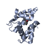
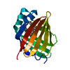
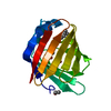
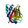
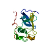
- PDB-1org: The crystal structure of a pheromone binding protein from the coc... -
+ Open data

- Basic information
| Entry | Database: PDB / ID: 1org | ||||||
|---|---|---|---|---|---|---|---|
| Title | The crystal structure of a pheromone binding protein from the cockroach Leucophaea maderae reveals a new mechanism of pheromone binding | ||||||
 Components Components | pheromone binding protein Insect pheromone-binding protein Insect pheromone-binding protein | ||||||
 Keywords Keywords | TRANSPORT PROTEIN / Pheromone binding Protein / APO-FORM / 6 ALPHA HELIX | ||||||
| Function / homology | Pheromone/general odorant binding protein domain / Pheromone/general odorant binding protein / PBP/GOBP family / Pheromone/general odorant binding protein superfamily / odorant binding / Recoverin; domain 1 / Orthogonal Bundle / Mainly Alpha / Pheromone binding protein Function and homology information Function and homology information | ||||||
| Biological species |  Leucophaea maderae (Madeira cockroach) Leucophaea maderae (Madeira cockroach) | ||||||
| Method | X-RAY DIFFRACTION / SYNCHROTRON / SAD / Resolution: 1.7 Å | ||||||
 Authors Authors | Lartigue, A. / Gruez, A. / Spinelli, S. / Riviere, S. / Brossut, R. / Tegoni, M. / Cambillau, C. | ||||||
 Citation Citation | Journal: J.Biol.Chem. / Year: 2003 Title: THE CRYSTAL STRUCTURE OF A COCKROACH PHEROMONE-BINDING PROTEIN SUGGESTS A NEW LIGAND BINDING AND RELEASE MECHANISM Authors: Lartigue, A. / Gruez, A. / Spinelli, S. / Riviere, S. / Brossut, R. / Tegoni, M. / Cambillau, C. | ||||||
| History |
|
- Structure visualization
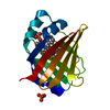
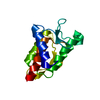
| Structure viewer | Molecule: MolmilJmol/JSmol |
|---|
- Downloads & links
-Download
| PDBx/mmCIF format | 1org.cif.gz | 57.3 KB | Display | PDBx/mmCIF format |
|---|---|---|---|---|
| PDB format | pdb1org.ent.gz | 46.1 KB | Display | PDB format |
| PDBx/mmJSON format | 1org.json.gz | Tree view | PDBx/mmJSON format | |
| Others |  Other downloads Other downloads |
-Validation report
| Arichive directory | https://data.pdbj.org/pub/pdb/validation_reports/or/1orgftp://data.pdbj.org/pub/pdb/validation_reports/or/1org | HTTPS FTP |
|---|
-Related structure data

-Links
 PDBj
PDBj- Assembly
| Deposited unit | 
| ||||||||
|---|---|---|---|---|---|---|---|---|---|
| 1 | 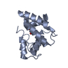
| ||||||||
| 2 | 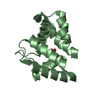
| ||||||||
| Unit cell |
|
-Components
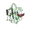
| #1: Protein | Insect pheromone-binding protein Mass: 13014.785 Da / Num. of mol.: 2 Source method: isolated from a genetically manipulated source Source: (gene. exp.) Leucophaea maderae (Madeira cockroach) / Plasmid: pET22b+ / Species (production host): Escherichia coli / Production host:  Escherichia coli BL21(DE3) (bacteria) / Strain (production host): BL21(DE3) / References: UniProt: Q8MTC1 Escherichia coli BL21(DE3) (bacteria) / Strain (production host): BL21(DE3) / References: UniProt: Q8MTC1#2: Chemical | Glycerol  Mass: 92.094 Da / Num. of mol.: 2 / Source method: obtained synthetically / Formula: C3H8O3 Mass: 92.094 Da / Num. of mol.: 2 / Source method: obtained synthetically / Formula: C3H8O3#3: Water | ChemComp-HOH / | Water Mass: 18.015 Da / Num. of mol.: 187 / Source method: isolated from a natural source / Formula: H2O Mass: 18.015 Da / Num. of mol.: 187 / Source method: isolated from a natural source / Formula: H2O |
|---|
-Experimental details
-Experiment
| Experiment | Method: X-RAY DIFFRACTION / Number of used crystals: 1 |
|---|
- Sample preparation
| Crystal | Density Matthews: 2.58 Å3/Da / Density % sol: 52 % |
|---|---|
| Crystal grow | Temperature: 293 K / Method: vapor diffusion / pH: 8 Details: Tris, PEG 2000, pH 8.0, VAPOR DIFFUSION, temperature 293K |
| Crystal grow | *PLUS Method: unknown / Details: Sulzenbacher, G., (2002) Acta Cryst., D58, 2109. |
-Data collection
| Diffraction | Mean temperature: 100 K |
|---|---|
| Diffraction source | Source: SYNCHROTRON / Site: ESRF  / Beamline: BM14 / Wavelength: 0.97887 Å / Beamline: BM14 / Wavelength: 0.97887 Å |
| Detector | Type: MARRESEARCH / Detector: CCD / Date: Jul 23, 2002 / Details: mirrors |
| Radiation | Monochromator: Mirrors / Protocol: SINGLE WAVELENGTH / Monochromatic (M) / Laue (L): M / Scattering type: x-ray |
| Radiation wavelength | Wavelength: 0.97887 Å / Relative weight: 1 |
| Reflection | Resolution: 1.7→25 Å / Num. all: 29653 / Num. obs: 29653 / % possible obs: 96 % / Observed criterion σ(F): 0 / Observed criterion σ(I): 0 / Rmerge(I) obs: 0.045 / Net I/σ(I): 8 |
| Reflection shell | Resolution: 1.7→1.79 Å / Redundancy: 5.8 % / Rmerge(I) obs: 0.045 / Mean I/σ(I) obs: 2.4 / % possible all: 94.8 |
| Reflection | *PLUS % possible obs: 95.7 % / Redundancy: 6 % |
| Reflection shell | *PLUS % possible obs: 94.8 % / Rmerge(I) obs: 0.274 |
- Processing
| Software |
| |||||||||||||||||||||||||||||||||||||||||||||||||||||||||||||||||||||||||||
|---|---|---|---|---|---|---|---|---|---|---|---|---|---|---|---|---|---|---|---|---|---|---|---|---|---|---|---|---|---|---|---|---|---|---|---|---|---|---|---|---|---|---|---|---|---|---|---|---|---|---|---|---|---|---|---|---|---|---|---|---|---|---|---|---|---|---|---|---|---|---|---|---|---|---|---|---|
| Refinement | Method to determine structure: SAD / Resolution: 1.7→20 Å / Cor.coef. Fo:Fc: 0.963 / Cor.coef. Fo:Fc free: 0.951 / SU B: 2.027 / SU ML: 0.067 / TLS residual ADP flag: LIKELY RESIDUAL / Cross valid method: THROUGHOUT / σ(F): 2.4 / ESU R: 0.106 / ESU R Free: 0.104 / Stereochemistry target values: MAXIMUM LIKELIHOOD / Details: HYDROGENS HAVE BEEN ADDED IN THE RIDING POSITIONS
| |||||||||||||||||||||||||||||||||||||||||||||||||||||||||||||||||||||||||||
| Solvent computation | Ion probe radii: 0.8 Å / Shrinkage radii: 0.8 Å / VDW probe radii: 1.4 Å / Solvent model: BABINET MODEL WITH MASK | |||||||||||||||||||||||||||||||||||||||||||||||||||||||||||||||||||||||||||
| Displacement parameters | Biso mean: 24.378 Å2
| |||||||||||||||||||||||||||||||||||||||||||||||||||||||||||||||||||||||||||
| Refinement step | Cycle: LAST / Resolution: 1.7→20 Å
| |||||||||||||||||||||||||||||||||||||||||||||||||||||||||||||||||||||||||||
| Refine LS restraints |
| |||||||||||||||||||||||||||||||||||||||||||||||||||||||||||||||||||||||||||
| LS refinement shell | Resolution: 1.7→1.744 Å / Total num. of bins used: 20 /
| |||||||||||||||||||||||||||||||||||||||||||||||||||||||||||||||||||||||||||
| Refinement TLS params. | Method: refined / Refine-ID: X-RAY DIFFRACTION
| |||||||||||||||||||||||||||||||||||||||||||||||||||||||||||||||||||||||||||
| Refinement TLS group |
| |||||||||||||||||||||||||||||||||||||||||||||||||||||||||||||||||||||||||||
| Refinement | *PLUS Highest resolution: 1.7 Å / Lowest resolution: 20 Å / Rfactor Rfree: 0.181 / Rfactor Rwork: 0.214 | |||||||||||||||||||||||||||||||||||||||||||||||||||||||||||||||||||||||||||
| Solvent computation | *PLUS | |||||||||||||||||||||||||||||||||||||||||||||||||||||||||||||||||||||||||||
| Displacement parameters | *PLUS | |||||||||||||||||||||||||||||||||||||||||||||||||||||||||||||||||||||||||||
| Refine LS restraints | *PLUS
| |||||||||||||||||||||||||||||||||||||||||||||||||||||||||||||||||||||||||||
| LS refinement shell | *PLUS Highest resolution: 1.7 Å / Lowest resolution: 1.74 Å |
