 Movie
Movie Controller


+ Open data

- Basic information
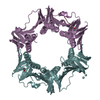
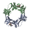
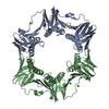
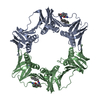
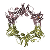
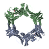
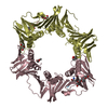
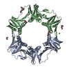
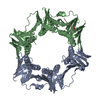
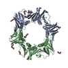
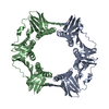
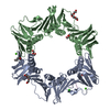
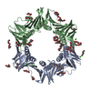
| Entry | Database: PDB / ID: 1ok7 | ||||||
|---|---|---|---|---|---|---|---|
| Title | A Conserved protein binding-site on Bacterial Sliding Clamps | ||||||
 Components Components |
| ||||||
 Keywords Keywords |  TRANSFERASE / DNA POLYMERASE IV / PEPTIDE INHIBITION / SLIDING CLAMP / TRANSLESION SYNTHESIS / DNA-DIRECTED DNA POLYMERASE / DNA REPLICATION TRANSFERASE / DNA POLYMERASE IV / PEPTIDE INHIBITION / SLIDING CLAMP / TRANSLESION SYNTHESIS / DNA-DIRECTED DNA POLYMERASE / DNA REPLICATION | ||||||
| Function / homology |  Function and homology information Function and homology informationHda-beta clamp complex / bacterial-type DNA replication / replication inhibiting complex / DNA polymerase III complex / replisome / SOS response / regulation of DNA-templated DNA replication initiation / DNA strand elongation involved in DNA replication / DNA synthesis involved in DNA repair / error-free translesion synthesis ...Hda-beta clamp complex / bacterial-type DNA replication / replication inhibiting complex / DNA polymerase III complex / replisome / SOS response / regulation of DNA-templated DNA replication initiation / DNA strand elongation involved in DNA replication / DNA synthesis involved in DNA repair / error-free translesion synthesis / error-prone translesion synthesis / negative regulation of DNA-templated DNA replication initiation / 3'-5' exonuclease activity / DNA-templated DNA replication / DNA replication / damaged DNA binding / DNA-directed DNA polymerase / DNA-directed DNA polymerase activity / DNA damage response / magnesium ion binding / protein homodimerization activity / DNA binding / identical protein binding / cytosol / cytoplasmSimilarity search - Function | ||||||
| Biological species |  ESCHERICHIA COLI (E. coli) ESCHERICHIA COLI (E. coli) | ||||||
| Method | X-RAY DIFFRACTION / SYNCHROTRON / MOLECULAR REPLACEMENT / Resolution: 1.65 Å | ||||||
 Authors Authors | Burnouf, D.Y. / Olieric, V. / Wagner, J. / Fujii, S. / Reinbolt, J. / Fuchs, R.P.P. / Dumas, P. | ||||||
 Citation Citation | Journal: J.Mol.Biol. / Year: 2004 Title: Structural and Biochemical Analysis of Sliding Clamp/Ligand Interactions Suggest a Competition between Replicative and Translesion DNA Polymerases Authors: Burnouf, D.Y. / Olieric, V. / Wagner, J. / Fujii, S. / Reinbolt, J. / Fuchs, R.P.P. / Dumas, P. #1: Journal: Cell(Cambridge,Mass.) / Year: 1992Title: Three-Dimensional Structure of the Beta Subunit of Escherichia Coli DNA Polymerase III Holoenzyme: A Sliding DNA Clamp Authors: Kong, X.-P. / Onrust, R. / O'Donnell, M. / Kuriyan, J. | ||||||
| History |
|
- Structure visualization
| Structure viewer | Molecule: MolmilJmol/JSmol |
|---|
- Downloads & links
-Download
| PDBx/mmCIF format | 1ok7.cif.gz | 163.1 KB | Display | PDBx/mmCIF format |
|---|---|---|---|---|
| PDB format | pdb1ok7.ent.gz | 128.5 KB | Display | PDB format |
| PDBx/mmJSON format | 1ok7.json.gz | Tree view | PDBx/mmJSON format | |
| Others |  Other downloads Other downloads |
-Validation report
| Arichive directory | https://data.pdbj.org/pub/pdb/validation_reports/ok/1ok7ftp://data.pdbj.org/pub/pdb/validation_reports/ok/1ok7 | HTTPS FTP |
|---|
-Related structure data
| Related structure data | 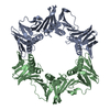 2polS S: Starting model for refinement |
|---|---|
| Similar structure data |


-Links
 PDBj
PDBj


- Assembly
| Deposited unit | 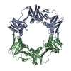
| ||||||||
|---|---|---|---|---|---|---|---|---|---|
| 1 |
| ||||||||
| Unit cell |
| ||||||||
| Noncrystallographic symmetry (NCS) | NCS oper: (Code: given Matrix: (0.18178, -0.82569, -0.53403), Vector : Details | PDB CONVENTION REQUIRES THAT THIS ENTRY BE CLASSIFIED AS TRIMERIC SINCE THE ASSEMBLY INCLUDES THREE CHAINS. HOWEVER CHAINS A AND B FORM A PHYSIOLOGICAL DIMER WHICH THEN INTERACTS WITH THE SMALLER CHAIN C. | |
-Components
| #1: Protein | DNA polymerase III holoenzyme / POLYMERASE / DNAN / B3701 / C4623 / Z5192 / ECS4636 Mass: 40630.508 Da / Num. of mol.: 2 Source method: isolated from a genetically manipulated source Source: (gene. exp.) ESCHERICHIA COLI (E. coli) / Description: DNAN GENE / Plasmid: PET15B / Production host: ESCHERICHIA COLI (E. coli) / Strain (production host): BL21(DE3)References: UniProt: P00583, UniProt: P0A988*PLUS, DNA-directed DNA polymerase#2: Protein/peptide | | / POL IVMass: 1826.185 Da / Num. of mol.: 1 / Fragment: C-TERMINUS OF DNA POL IV RESIDUES 336-351 / Source method: obtained synthetically / Details: C-TERMINUS (16 RESIDUES) OF DNA POL IV E.COLI / Source: (synth.) ESCHERICHIA COLI (E. coli) / References: UniProt: Q47155, DNA-directed DNA polymerase#3: Water | ChemComp-HOH / | Water Mass: 18.015 Da / Num. of mol.: 460 / Source method: isolated from a natural source / Formula: H2O Mass: 18.015 Da / Num. of mol.: 460 / Source method: isolated from a natural source / Formula: H2OCompound details | CATALYTIC ACTIVITY: N DEOXYNUCLE | Sequence details | DISORDERED LOOP (A19 - A26) WAS BUILT ONLY ON A STEREOCHEMICAL BASIS. ARG C10 WAS BUILT IN A RATHER ...DISORDERED | |
|---|
-Experimental details
-Experiment
| Experiment | Method: X-RAY DIFFRACTION / Number of used crystals: 1 |
|---|
- Sample preparation
| Crystal | Density Matthews: 2.32 Å3/Da / Density % sol: 45 % |
|---|---|
| Crystal grow | pH: 6 Details: DROPS:0.92 ML OF PROTEIN AT 34.2 MG/ML, 1.89 ML OF P16 AT 1.1 MG/ML, 1 ML OF 2X RESERVOIR SOLUTION. RESERVOIR: 0.1 M MES PH 6.0, 0.1M CACL2 AND 30% PEG 400 |
-Data collection
| Diffraction | Mean temperature: 100 K |
|---|---|
| Diffraction source | Source: SYNCHROTRON / Site: ESRF  / Beamline: ID14-4 / Wavelength: 0.93922 / Beamline: ID14-4 / Wavelength: 0.93922 |
| Detector | Type: ADSC CCD / Detector: CCD / Date: Apr 15, 2002 / Details: MIRRORS |
| Radiation | Protocol: SINGLE WAVELENGTH / Monochromatic (M) / Laue (L): M / Scattering type: x-ray |
| Radiation wavelength | Wavelength: 0.93922 Å / Relative weight: 1 |
| Reflection | Resolution: 1.65→20 Å / Num. obs: 85999 / % possible obs: 96.7 % / Redundancy: 2.7 % / Biso Wilson estimate: 19.8 Å2 / Rmerge(I) obs: 0.051 / Net I/σ(I): 12.51 |
| Reflection shell | Resolution: 1.65→1.75 Å / Redundancy: 2.7 % / Rmerge(I) obs: 0.178 / Mean I/σ(I) obs: 5.23 / % possible all: 95.6 |
- Processing
| Software |
| ||||||||||||||||||||||||||||||||||||||||||||||||||||||||||||||||||||||||||||||||
|---|---|---|---|---|---|---|---|---|---|---|---|---|---|---|---|---|---|---|---|---|---|---|---|---|---|---|---|---|---|---|---|---|---|---|---|---|---|---|---|---|---|---|---|---|---|---|---|---|---|---|---|---|---|---|---|---|---|---|---|---|---|---|---|---|---|---|---|---|---|---|---|---|---|---|---|---|---|---|---|---|---|
| Refinement | Method to determine structure: MOLECULAR REPLACEMENT Starting model: PDB ENTRY 2POL Resolution: 1.65→20 Å / Rfactor Rfree error: 0.004 / Data cutoff high absF: 0 / Isotropic thermal model: RESTRAINED / Cross valid method: THROUGHOUT / σ(F): 0
| ||||||||||||||||||||||||||||||||||||||||||||||||||||||||||||||||||||||||||||||||
| Solvent computation | Solvent model: FLAT MODEL / Bsol: 48.396 Å2 / ksol: 0.368559 e/Å3 | ||||||||||||||||||||||||||||||||||||||||||||||||||||||||||||||||||||||||||||||||
| Displacement parameters | Biso mean: 23.9 Å2
| ||||||||||||||||||||||||||||||||||||||||||||||||||||||||||||||||||||||||||||||||
| Refine analyze |
| ||||||||||||||||||||||||||||||||||||||||||||||||||||||||||||||||||||||||||||||||
| Refinement step | Cycle: LAST / Resolution: 1.65→20 Å
| ||||||||||||||||||||||||||||||||||||||||||||||||||||||||||||||||||||||||||||||||
| Refine LS restraints |
| ||||||||||||||||||||||||||||||||||||||||||||||||||||||||||||||||||||||||||||||||
| Refine LS restraints NCS | NCS model details: UNRESTRAINED | ||||||||||||||||||||||||||||||||||||||||||||||||||||||||||||||||||||||||||||||||
| LS refinement shell | Resolution: 1.65→1.75 Å / Rfactor Rfree error: 0.011 / Total num. of bins used: 6
|
