 Movie
Movie Controller


+ Open data

- Basic information
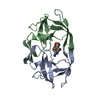
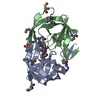
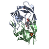
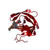
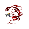
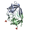
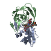
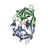
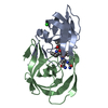
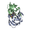
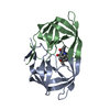
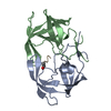
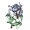
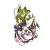
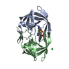
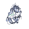
| Entry | Database: PDB / ID: 1ody | ||||||
|---|---|---|---|---|---|---|---|
| Title | HIV-1 PROTEASE COMPLEXED WITH AN INHIBITOR LP-130 | ||||||
 Components Components | HIV-1 PROTEASE | ||||||
 Keywords Keywords | ASPARTIC PROTEASE / RETROPEPSIN / RETROVIRUS / HIV / AIDS | ||||||
| Function / homology |  Function and homology informationintegrase activity / Integration of viral DNA into host genomic DNA / Autointegration results in viral DNA circles / Minus-strand DNA synthesis / Plus-strand DNA synthesis / Uncoating of the HIV Virion / 2-LTR circle formation / Early Phase of HIV Life Cycle / Vpr-mediated nuclear import of PICs / Integration of provirus ...integrase activity / Integration of viral DNA into host genomic DNA / Autointegration results in viral DNA circles / Minus-strand DNA synthesis / Plus-strand DNA synthesis / Uncoating of the HIV Virion / 2-LTR circle formation / Early Phase of HIV Life Cycle / Vpr-mediated nuclear import of PICs / Integration of provirus / APOBEC3G mediated resistance to HIV-1 infection / Binding and entry of HIV virion / viral life cycle / Assembly Of The HIV Virion / HIV-1 retropepsin / : / retroviral ribonuclease H / Budding and maturation of HIV virion / exoribonuclease H / : / exoribonuclease H activity / protein processing / host multivesicular body / RNA-directed DNA polymerase / viral genome integration into host DNA / viral penetration into host nucleus / establishment of integrated proviral latency / RNA-directed DNA polymerase activity / Transferases; Transferring phosphorus-containing groups; Nucleotidyltransferases / RNA-DNA hybrid ribonuclease activity / peptidase activity / viral nucleocapsid / DNA recombination / Hydrolases; Acting on ester bonds / DNA-directed DNA polymerase / aspartic-type endopeptidase activity / DNA-directed DNA polymerase activity / symbiont entry into host cell / symbiont-mediated suppression of host gene expression / lipid binding / host cell nucleus / host cell plasma membrane / virion membrane / structural molecule activity / DNA binding / RNA binding / zinc ion binding / membrane / identical protein binding Function and homology informationintegrase activity / Integration of viral DNA into host genomic DNA / Autointegration results in viral DNA circles / Minus-strand DNA synthesis / Plus-strand DNA synthesis / Uncoating of the HIV Virion / 2-LTR circle formation / Early Phase of HIV Life Cycle / Vpr-mediated nuclear import of PICs / Integration of provirus ...integrase activity / Integration of viral DNA into host genomic DNA / Autointegration results in viral DNA circles / Minus-strand DNA synthesis / Plus-strand DNA synthesis / Uncoating of the HIV Virion / 2-LTR circle formation / Early Phase of HIV Life Cycle / Vpr-mediated nuclear import of PICs / Integration of provirus / APOBEC3G mediated resistance to HIV-1 infection / Binding and entry of HIV virion / viral life cycle / Assembly Of The HIV Virion / HIV-1 retropepsin / : / retroviral ribonuclease H / Budding and maturation of HIV virion / exoribonuclease H / : / exoribonuclease H activity / protein processing / host multivesicular body / RNA-directed DNA polymerase / viral genome integration into host DNA / viral penetration into host nucleus / establishment of integrated proviral latency / RNA-directed DNA polymerase activity / Transferases; Transferring phosphorus-containing groups; Nucleotidyltransferases / RNA-DNA hybrid ribonuclease activity / peptidase activity / viral nucleocapsid / DNA recombination / Hydrolases; Acting on ester bonds / DNA-directed DNA polymerase / aspartic-type endopeptidase activity / DNA-directed DNA polymerase activity / symbiont entry into host cell / symbiont-mediated suppression of host gene expression / lipid binding / host cell nucleus / host cell plasma membrane / virion membrane / structural molecule activity / DNA binding / RNA binding / zinc ion binding / membrane / identical protein bindingSimilarity search - Function | ||||||
| Biological species |  HIV-1 M:B_HXB2R (virus) HIV-1 M:B_HXB2R (virus) | ||||||
| Method | X-RAY DIFFRACTION / RIGID BODY REFINEMENT / Resolution: 2 Å | ||||||
 Authors Authors | Kervinen, J. / Lubkowski, J. / Zdanov, A. / Wlodawer, A. / Gustchina, A. | ||||||
 Citation Citation | Journal: Protein Sci. / Year: 1998 Title: Toward a universal inhibitor of retroviral proteases: comparative analysis of the interactions of LP-130 complexed with proteases from HIV-1, FIV, and EIAV. Authors: Kervinen, J. / Lubkowski, J. / Zdanov, A. / Bhatt, D. / Dunn, B.M. / Hui, K.Y. / Powell, D.J. / Kay, J. / Wlodawer, A. / Gustchina, A. | ||||||
| History |
|
- Structure visualization
| Structure viewer | Molecule: MolmilJmol/JSmol |
|---|
- Downloads & links
-Download
| PDBx/mmCIF format | 1ody.cif.gz | 50.7 KB | Display | PDBx/mmCIF format |
|---|---|---|---|---|
| PDB format | pdb1ody.ent.gz | 39.8 KB | Display | PDB format |
| PDBx/mmJSON format | 1ody.json.gz | Tree view | PDBx/mmJSON format | |
| Others |  Other downloads Other downloads |
-Validation report
| Arichive directory | https://data.pdbj.org/pub/pdb/validation_reports/od/1odyftp://data.pdbj.org/pub/pdb/validation_reports/od/1ody | HTTPS FTP |
|---|
-Related structure data
| Related structure data | 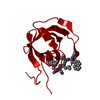 2fmbC 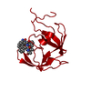 4fivC 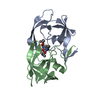 1odxS S: Starting model for refinement C: citing same article ( |
|---|---|
| Similar structure data |

-Links
 PDBj
PDBj


- Assembly
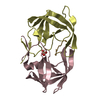
| Deposited unit | 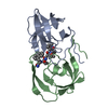
| ||||||||
|---|---|---|---|---|---|---|---|---|---|
| 1 |
| ||||||||
| Unit cell |
|
-Components
| #1: Protein | / HIV-1 PR / RETROPEPSIN Mass: 10703.613 Da / Num. of mol.: 2 Source method: isolated from a genetically manipulated source Source: (gene. exp.) HIV-1 M:B_HXB2R (virus) / Genus: Lentivirus / Species: Human immunodeficiency virus 1Subtypes of HIV / Production host:  Escherichia coli (E. coli) / Strain (production host): HXB2 Escherichia coli (E. coli) / Strain (production host): HXB2References: PIR: S63716, UniProt: P04585*PLUS, HIV-1 retropepsin#2: Chemical | ChemComp-LP1 / | 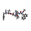  Mass: 794.978 Da / Num. of mol.: 1 / Source method: obtained synthetically / Formula: C45H58N6O7 Mass: 794.978 Da / Num. of mol.: 1 / Source method: obtained synthetically / Formula: C45H58N6O7#3: Water | ChemComp-HOH / | Water Mass: 18.015 Da / Num. of mol.: 145 / Source method: isolated from a natural source / Formula: H2O Mass: 18.015 Da / Num. of mol.: 145 / Source method: isolated from a natural source / Formula: H2ONonpolymer details | LP-130 (HET ID LP1): ACE-NAL-VAL-STA-ABU-NAL-NH2 WHERE NAL IS NAPHTHYLALANINE, STA IS STATINE, AND ...LP-130 (HET ID LP1): ACE-NAL-VAL-STA-ABU-NAL-NH2 WHERE NAL IS NAPHTHYLAL | |
|---|
-Experimental details
-Experiment
| Experiment | Method: X-RAY DIFFRACTION / Number of used crystals: 1 |
|---|
- Sample preparation
| Crystal | Density Matthews: 2.22 Å3/Da / Density % sol: 44.62 % | ||||||||||||||||||||||||||||||
|---|---|---|---|---|---|---|---|---|---|---|---|---|---|---|---|---|---|---|---|---|---|---|---|---|---|---|---|---|---|---|---|
| Crystal grow | pH: 6.2 / Details: pH 6.2 | ||||||||||||||||||||||||||||||
| Crystal grow | *PLUS Method: vapor diffusion, hanging drop | ||||||||||||||||||||||||||||||
| Components of the solutions | *PLUS
|
-Data collection
| Diffraction | Mean temperature: 293 K |
|---|---|
| Diffraction source | Source: ROTATING ANODE / Type: ENRAF-NONIUS FR591 / Wavelength: 1.5418 |
| Detector | Type: MACSCIENCE / Detector: IMAGE PLATE / Date: Jul 11, 1997 / Details: MIRRORS |
| Radiation | Monochromator: NI FILTER / Monochromatic (M) / Laue (L): M / Scattering type: x-ray |
| Radiation wavelength | Wavelength: 1.5418 Å / Relative weight: 1 |
| Reflection | Resolution: 2→20 Å / Num. obs: 12082 / % possible obs: 95 % / Observed criterion σ(I): 1 / Redundancy: 4.7 % / Rmerge(I) obs: 0.067 |
| Reflection shell | Resolution: 2→2.07 Å / Rmerge(I) obs: 0.429 / % possible all: 85.1 |
| Reflection shell | *PLUS % possible obs: 85.1 % |
- Processing
| Software |
| ||||||||||||||||||||||||||||||||||||||||||||||||||||||||||||||||||||||||||||||||||||
|---|---|---|---|---|---|---|---|---|---|---|---|---|---|---|---|---|---|---|---|---|---|---|---|---|---|---|---|---|---|---|---|---|---|---|---|---|---|---|---|---|---|---|---|---|---|---|---|---|---|---|---|---|---|---|---|---|---|---|---|---|---|---|---|---|---|---|---|---|---|---|---|---|---|---|---|---|---|---|---|---|---|---|---|---|---|
| Refinement | Method to determine structure: RIGID BODY REFINEMENT Starting model: PDB ENTRY 1ODX Resolution: 2→8 Å / σ(F): 3 /
| ||||||||||||||||||||||||||||||||||||||||||||||||||||||||||||||||||||||||||||||||||||
| Refinement step | Cycle: LAST / Resolution: 2→8 Å
| ||||||||||||||||||||||||||||||||||||||||||||||||||||||||||||||||||||||||||||||||||||
| Refine LS restraints |
|
